. 2020 Nov 3:9:e61245.
doi: 10.7554/eLife.61245.

Atg43 tethers isolation membranes to mitochondria to promote starvation-induced mitophagy in fission yeast

Affiliations

Atg43 tethers isolation membranes to mitochondria to promote starvation-induced mitophagy in fission yeast

Tomoyuki Fukuda et al. Elife. .

Abstract

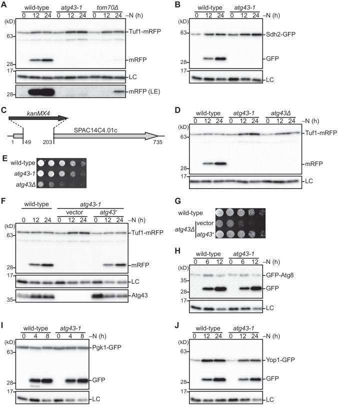
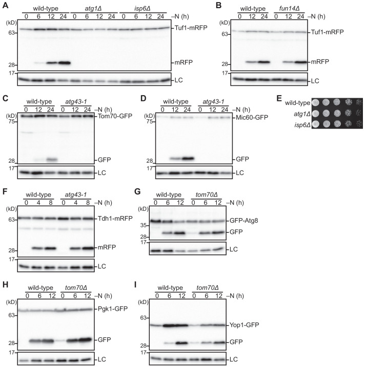
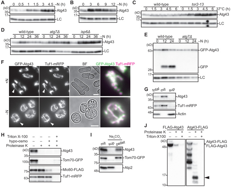
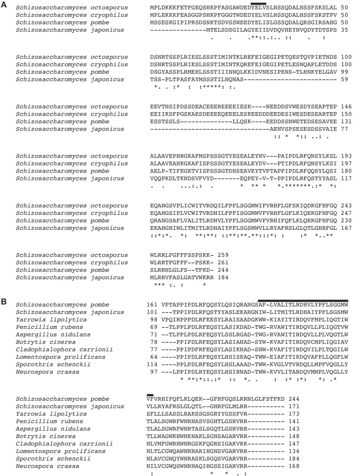
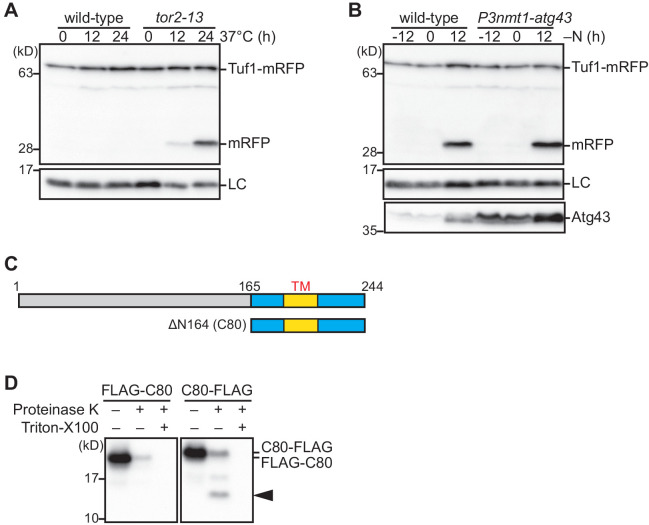
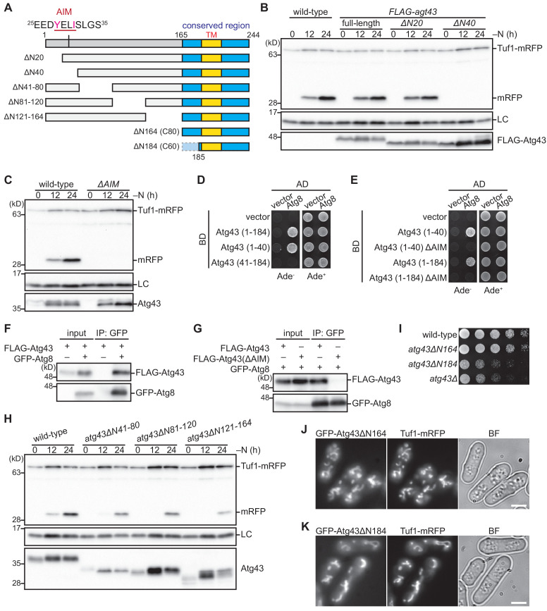
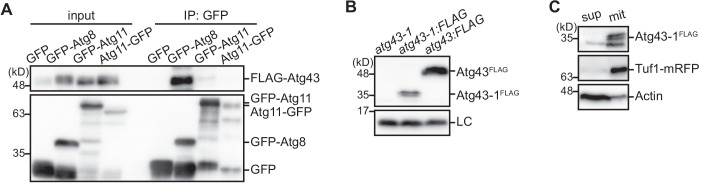
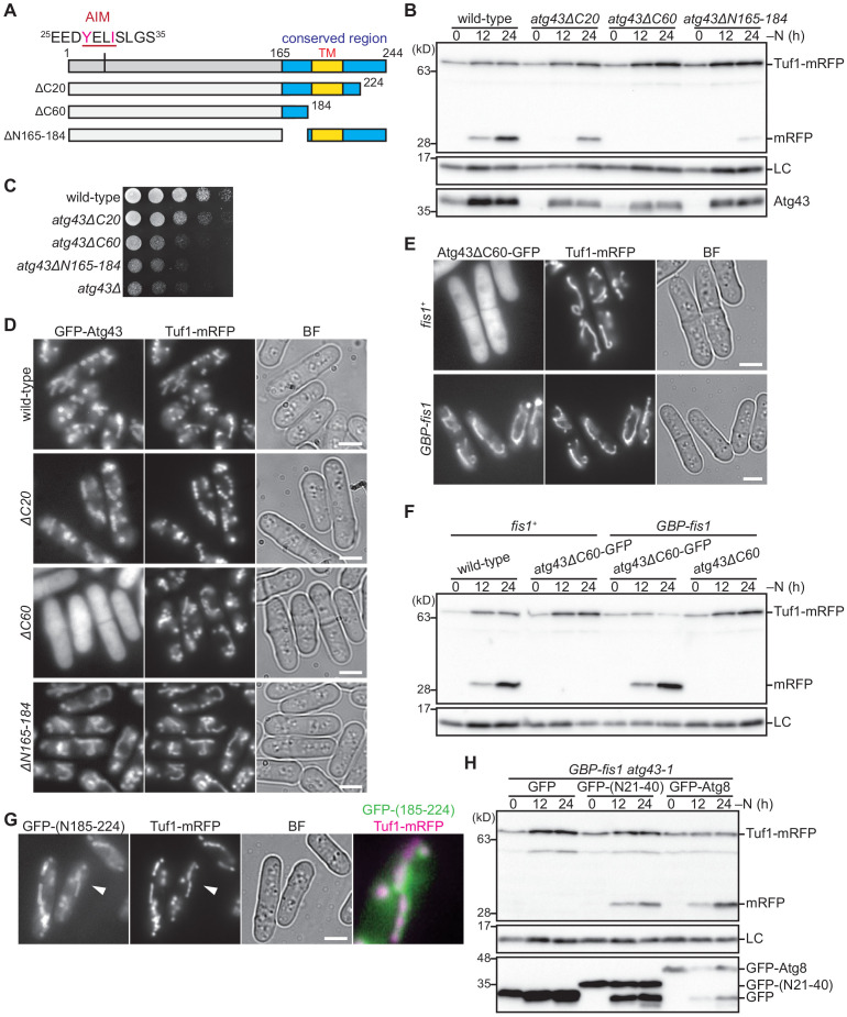
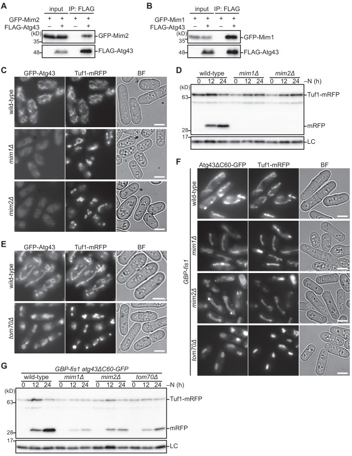
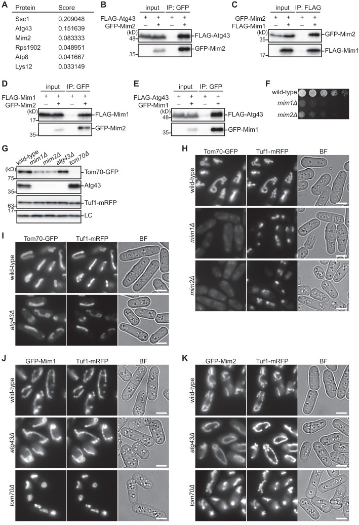
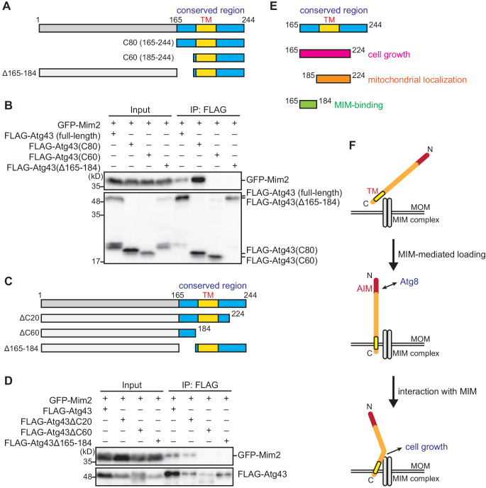
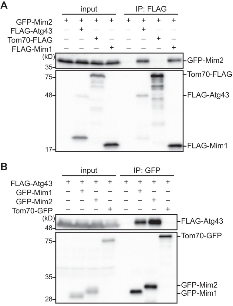
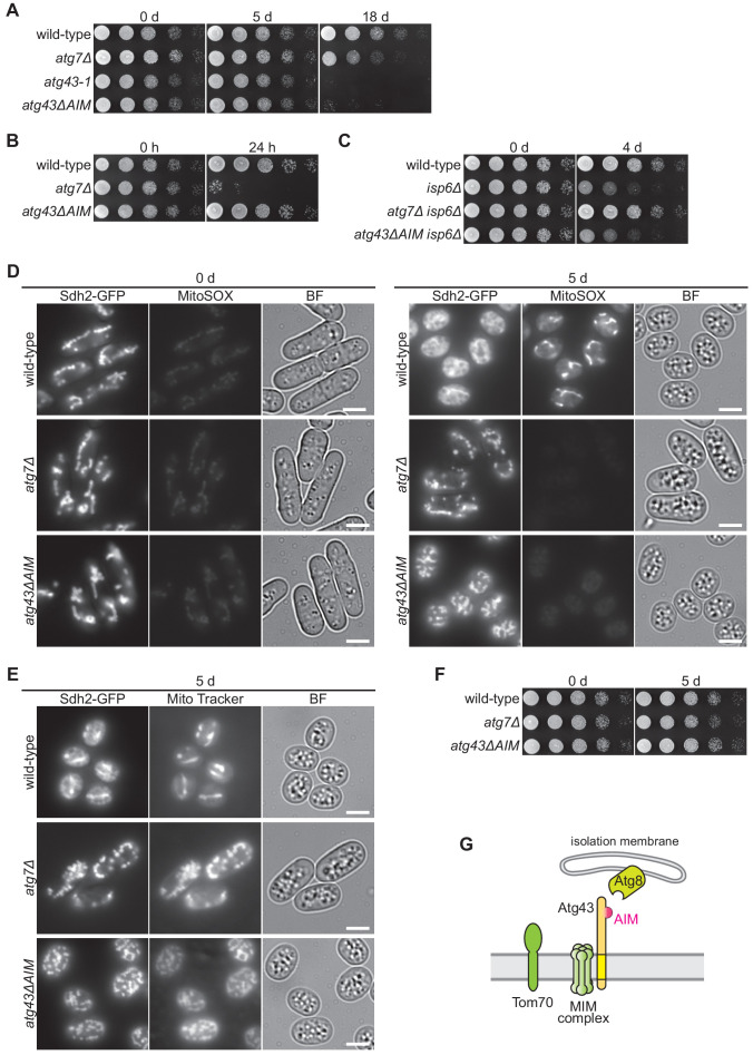
Degradation of mitochondria through mitophagy contributes to the maintenance of mitochondrial function. In this study, we identified that Atg43, a mitochondrial outer membrane protein, serves as a mitophagy receptor in the model organism Schizosaccharomyces pombe to promote the selective degradation of mitochondria. Atg43 contains an Atg8-family-interacting motif essential for mitophagy. Forced recruitment of Atg8 to mitochondria restores mitophagy in Atg43-deficient cells, suggesting that Atg43 tethers expanding isolation membranes to mitochondria. We found that the mitochondrial import factors, including the Mim1-Mim2 complex and Tom70, are crucial for mitophagy. Artificial mitochondrial loading of Atg43 bypasses the requirement of the import factors, suggesting that they contribute to mitophagy through Atg43. Atg43 not only maintains growth ability during starvation but also facilitates vegetative growth through its mitophagy-independent function. Thus, Atg43 is a useful model to study the mechanism and physiological roles, as well as the origin and evolution, of mitophagy in eukaryotes.

Keywords: Atg43; MIM complex; S. pombe; autophagy; cell biology; mitochondria; mitophagy; receptor.


Conflict of interest statement

TF, YE, TS, KF, SY, KI, DK, YY, TK No competing interests declared

Figures

Figure 1.
Figure 1.. Atg43 is required for starvation-induced mitochondrial degradation in S. pombe.
(A, D) The indicated strains expressing Tuf1-mRFP were grown in EMM and shifted to the same medium without a nitrogen source (EMM–N). Cells were collected at the indicated time points after nitrogen starvation was initiated. Tuf1-mRFP processing was monitored by immunoblotting. Lower panel, long exposure (LE). (B) The indicated strains expressing Sdh2-GFP were collected at the indicated time points after shifting to nitrogen starvation medium. The processing of Sdh2-GFP was monitored by immunoblotting. (C) Schematic representation of the atg43-1 allele. The protein-coding region is depicted by a gray arrow. The nucleotide region that is deleted by the replacement with the kanMX4 gene (black arrow) is shown. (E, G) The indicated strains were grown in EMM, and their serial dilutions were spotted onto solid YES medium for the growth assay. (F) The protein-coding region of the atg43+ gene was cloned into an integration vector to express atg43+ under the control of the moderate adh31 promoter. The atg43-1 mutants integrated with the atg43+ gene (atg43+) or an empty vector (vector), as well as a wild-type strain, were collected at the indicated time points after induction of nitrogen starvation and were subsequently subjected to immunoblotting. (H–J) The indicated strains expressing GFP-Atg8, Pgk1-GFP, or Yop1-GFP were collected at the indicated time points after induction of nitrogen starvation. Degradation of each substrate was monitored by immunoblotting. Histone H3 was used as a loading control (LC) for immunoblotting.
Figure 1—figure supplement 1.
Figure 1—figure supplement 1.. Mitochondrial proteins are degraded by autophagy dependently on Atg43 and Tom70.
(A–D) The processing of tagged mitochondrial proteins depends on mitochondrial degradation by autophagy. Wild-type and the indicated mutant cells, which express Tuf1-mRFP (A and B), Tom70-GFP (C), or Mic60-GFP (D), were grown in EMM and shifted to EMM–N for nitrogen starvation. Cells were collected at the indicated time points after induction of nitrogen starvation, followed by immunoblotting. Histone H3 was used as a loading control (LC). (E) Autophagy does not affect vegetative growth. The indicated strains were grown in EMM, and their serial dilutions were spotted onto solid YES medium for the growth assay. (F–I) Atg43 and Tom70 are mitophagy-specific proteins. Wild-type and the indicated mutant cells expressing Tdh1-mRFP (F), GFP-Atg8 (G), Pgk1-GFP (H), or Yop1-GFP (I) were grown in EMM and shifted to EMM–N for nitrogen starvation. Cells were collected at the indicated time points after induction of nitrogen starvation and were used for immunoblotting.
Figure 2.
Figure 2.. Atg43 is a transmembrane protein localized to the MOM.
(A, B) Wild-type cells were grown in EMM and shifted to EMM–N for nitrogen starvation. Cells were collected at the indicated time points after shifting to nitrogen starvation medium and were then subjected to immunoblotting. (C) The indicated strains were grown in EMM at 26°C. The temperature was then shifted to 37°C to inactivate TORC1. Cells were collected at the indicated time points after the temperature shift and were used for immunoblotting. (D, E) The indicated strains were collected at the indicated time points after shifting to nitrogen starvation medium and were used for immunoblotting. (F) Cells expressing GFP-Atg43 and Tuf1-mRFP were grown in EMM (+N) or cultured in EMM–N for 12 hr (–N) for microscopy. A magnified view of the indicated area is shown in the right panel. Scale bars represent 5 µm. BF, bright-field image. (G) Fractionation was conducted using cells expressing Tuf1-mRFP. The total cell homogenate (total) was fractionated by centrifugation to obtain a mitochondria-enriched pellet (mit) and supernatant (sup). Tuf1-mRFP and actin were detected as markers of the mitochondria and cytosol, respectively. (H) A mitochondrial fraction was prepared from cells expressing Tom70-GFP, Mic60-FLAG, and Tuf1-mRFP, treated with (+) or without (–) proteinase K, under different conditions. Hypo-osmotic swelling resulted in rupture of the MOM and the detergent Triton X-100 lysed mitochondria. Tom70-GFP, Mic60-FLAG, and Tuf1-mRFP were detected as markers of the mitochondrial outer membrane, inner membrane, and matrix, respectively. (I) The mitochondrial fraction (mit) was prepared from cells expressing Tom70-GFP, treated with sodium carbonate, and separated into the soluble supernatant (sup) and membrane pellet (pellet) by centrifugation. Tom70-GFP and Atp2 were detected as markers for integral and peripheral membrane proteins, respectively. (J) The mitochondrial fraction was prepared from cells expressing N-terminal or C-terminal FLAG-tagged Atg43 (FLAG-Atg43 or Atg43-FLAG, respectively), followed by treatment with (+) or without (–) proteinase K. The Triton X-100 detergent lysed mitochondria. The proteinase-resistant band is indicated by the arrowhead. Histone H3 was used as a loading control (LC) for immunoblotting.
Figure 2—figure supplement 1.
Figure 2—figure supplement 1.. The C-terminal region of Atg43 is conserved.
(A) A multiple sequence alignment of Atg43 proteins in the Schizosaccharomyces species. The alignment was produced using ClustalW and modified manually. The N-terminal AIM is indicated. (B) Alignment of fungal sequences similar to the C-terminal region of Atg43. The alignment was produced using ClustalW and modified manually. The predicted C-terminal transmembrane domain is indicated.
Figure 2—figure supplement 2.
Figure 2—figure supplement 2.. Atg43 is a transmembrane protein that is expressed during nitrogen starvation.
(A) The indicated strains were grown in EMM and the temperature was shifted to 37°C to inactivate TORC1. Cells were collected at the indicated time points after the temperature shift and were used for immunoblotting. (B) Wild-type cells and cells expressing Atg43 under the thiamine-repressible nmt1 promoter were grown in EMM containing thiamine for moderate expression. Cells were collected at the indicated time points after shifting to nitrogen starvation medium and were used for immunoblotting. Histone H3 was used as a loading control (LC). (C) Schematic representation of the structure of Atg43. The region conserved among fungi is highlighted in blue. A C-terminal predicted transmembrane domain (TM) is indicated. (D) Cells expressing the C-terminal 80 aa of Atg43 (C80) tagged with FLAG at the N-terminus (FLAG-C80) or C-terminus (C80-FLAG) were grown in YES. The mitochondrial fraction was prepared and treated with (+) or without (–) proteinase K. The detergent Triton X-100 lysed mitochondria. The proteinase-resistant band is indicated by an arrowhead.
Figure 3.
Figure 3.. Atg43 contains an AIM that is essential for mitophagy.
(A) Schematic representation of N-terminal deleted forms of Atg43. The region conserved among fungi is highlighted in blue. The N-terminal AIM and C-terminal predicted transmembrane domain (TM) are indicated. (B) Wild-type cells and strains expressing FLAG-tagged Atg43, with or without N-terminal truncation, under the thiamine-repressible nmt1 promoter, were grown in EMM containing thiamine for moderate expression. Cells were collected at the indicated time points after shifting to nitrogen starvation medium and were subsequently used for immunoblotting. (C) Cells expressing wild-type and AIM-mutated (ΔAIM) Atg43 from the native gene locus were collected at the indicated time points after shifting to nitrogen starvation medium and subjected to immunoblotting. (D, E) A yeast two-hybrid assay between activation domain (AD)-fused Atg8 and DNA-binding domain (BD)-fused Atg43 fragments, without or with mutations in the N-terminal AIM (ΔAIM). (F, G) Crude cell lysates (input) were prepared from cells with (+) or without (–) expression of GFP-fused Atg8 and FLAG-tagged Atg43 or AIM-mutated Atg43 (ΔAIM). Anti-GFP immunoprecipitants (IP: GFP) were analyzed by immunoblotting. Cells were collected 12 hr after shifting to nitrogen starvation medium and were subjected to immunoprecipitation. (H) Cells expressing wild-type and deleted forms of Atg43 from the native gene locus were collected at the indicated time points after shifting to nitrogen starvation medium and were subsequently used for immunoblotting. (I) The indicated strains were grown in EMM, and their serial dilutions were spotted onto solid YES medium for the growth assay. (J, K) Cells expressing N-terminal truncated forms of Atg43 tagged with GFP were grown in EMM for microscopy. Tuf1-mRFP was detected as a mitochondrial marker. Scale bars represent 5 µm. BF, bright-field image. Histone H3 was used as a loading control (LC) for immunoblotting.
Figure 3—figure supplement 1.
Figure 3—figure supplement 1.. The N-terminal region of Atg43 interacts with Atg8 and is dispensable for the mitochondrial localization.
(A) Immunoprecipitation experiments were conducted using crude cell lysates (input) prepared from FLAG-Atg43-expressing cells with co-expression of the indicated proteins. Cells were collected 12 hr after induction of nitrogen starvation and subjected to immunoprecipitation. (B) The atg43-1 allele was chromosomally tagged with FLAG (atg43-1:FLAG) and the expressed protein was detected by immunoblotting in parallel with the untagged atg43-1 (atg43-1) and tagged wild-type (atg43:FLAG) strains. Histone H3 was used as a loading control (LC). (C) Fractionation was conducted using atg43-1:FLAG cells. The cell homogenate was fractionated by centrifugation to obtain a mitochondria-enriched pellet (mit) and supernatant (sup). Tuf1-mRFP and actin were detected as markers of the mitochondria and cytosol, respectively.
Figure 4.
Figure 4.. The C-terminal region of Atg43 that contains a predicted transmembrane domain is required for mitochondrial localization.
(A) Schematic representation of C-terminal deleted forms of Atg43. The region conserved among fungi is highlighted in blue. The N-terminal AIM and predicted C-terminal transmembrane domain (TM) are indicated. (B) Cells expressing wild-type and C-terminal deleted forms of Atg43 from the native gene locus were collected at the indicated time points after shifting to nitrogen starvation medium and were subjected to immunoblotting. Each strain contained an integration vector expressing the mitophagy-defective form of Atg43, that lacks the 164 N-terminal aa, to maintain a normal growth rate. (C) The indicated strains were grown in EMM, and their serial dilutions were spotted onto solid YES medium for a growth assay. (D) Cells expressing wild-type and C-terminal truncated forms of GFP-tagged Atg43 were grown in EMM for microscopy. Tuf1-mRFP was detected as a mitochondrial marker. (E) Wild-type (fis1+) and GBP-fis1 cells co-expressing GFP-tagged Atg43 with a 60 aa deletion in the C-terminus and Tuf1-mRFP were grown in EMM for microscopy. (F) Wild-type (fis1+) and GBP-fis1 cells expressing full-length (wild-type) or C-terminal truncated Atg43 (Atg40ΔC60) with or without GFP fusion were collected at the indicated time points after shifting to nitrogen starvation medium. Cells were then used for immunoblotting. (G) Cells expressing the GFP-fused aa 185–224 of Atg43 and Tuf1-mRFP were grown in EMM for microscopy. A magnified view of the indicated area is shown in the right panel. (H) Mitophagy-defective atg43-1 cells with GBP-fused Fis1 were transformed to express GFP, GFP-fused aa 21–40 of Atg43, or GFP-fused Atg8. The indicated strains were collected at the indicated time points after shifting to nitrogen starvation medium and used for immunoblotting. Histone H3 was used as a loading control (LC) for immunoblotting. Scale bars represent 5 µm. BF, bright-field image.
Figure 5.
Figure 5.. The MIM complex facilitates mitophagy through the loading of Atg43 to the MOM.
(A, B) Crude cell lysates (input) were prepared from cells co-expressing GFP-fused Mim2 or Mim1 with (+) or without (–) FLAG-tagged Atg43. Anti-FLAG immunoprecipitants (IP: FLAG) were analyzed by immunoblotting. (C, E) The indicated strains co-expressing GFP-Atg43 and Tuf1-mRFP were grown in EMM for microscopy. (D) The indicated strains expressing Tuf1-mRFP were collected at the indicated time points after shifting to nitrogen starvation medium and were used for immunoblotting. (F) The indicated strains co-expressing a GFP-fused C-terminal truncated form of Atg43 (Atg43ΔC60-GFP) and Tuf1-mRFP were grown in EMM for microscopy. (G) Wild-type, mim1Δ, mim2Δ, and tom70Δ cells co-expressing GBP-fused Fis1 and GFP-fused Atg43 lacking the 60 C-terminal aa (Atg43ΔC60-GFP) were collected at the indicated time points after shifting to nitrogen starvation medium. Cells were then used for immunoblotting. Histone H3 was used as a loading control (LC) for immunoblotting. Scale bars represent 5 µm. BF, bright-field image.
Figure 5—figure supplement 1.
Figure 5—figure supplement 1.. The MIM complex facilitates the mitochondrial localization of Atg43 and Tom70.
(A) Identification of Mim2 as an Atg43-binding protein. Mass spectrometric analysis of proteins co-purified with the 80 C-terminal aa of Atg43 with a FLAG tag was conducted. Wild-type cells expressing untagged Atg43 were used as a negative control. The value (score) represents the relative protein abundance as expressed by spectral abundance factor (SAF). Mim2 was identified along with 37 peptide-spectrum matches, at a targeted false discovery rate (FDR) of 1.0% at the peptide level. (B–E) Atg43 interacts with the MIM complex. Immunoprecipitation experiments were conducted using crude cell lysates (input) prepared from cells with (+) or without (–) expression of the indicated proteins. Anti-FLAG (IP: FLAG) and anti-GFP (IP: GFP) immunoprecipitants were analyzed by immunoblotting. (F) The indicated strains were grown in EMM, and their serial dilutions were spotted onto solid YES medium for the growth assay. (G) Atg43 is unstable in the absence of the MIM complex. The indicated strains co-expressing Tom70-GFP and Tuf1-mRFP were grown in EMM for immunoblotting. Histone H3 was used as a loading control (LC). (H, I) Mitochondrial localization of Tom70 requires the MIM complex but does not require Atg43. The indicated strains co-expressing GFP-tagged Tom70 with Tuf1-mRFP were grown in EMM for microscopy. (J, K) Mitochondrial localization of the MIM complex is independent of Atg43 and Tom70. The indicated strains co-expressing Tuf1-mRFP with GFP-tagged Mim1 (J) or Mim2 (K) were grown in EMM for microscopy. Scale bars denote 5 µm. BF, bright-field image.
Figure 6.
Figure 6.. C-terminal region of Atg43 is required for its interaction with the MIM complex.
(A, C) Schematic representation of deleted forms of Atg43. The region conserved among fungi is highlighted in blue. The C-terminal predicted transmembrane domain (TM) is also indicated. (B, D) Crude cell lysates (input) were prepared from cells co-expressing GFP-fused Mim2 and FLAG-tagged Atg43 with or without deletion. Anti-FLAG immunoprecipitants (IP: FLAG) were analyzed by immunoblotting. (E) Schematic representation of the C-terminal region of Atg43. (F) Schematic model describing the loading and function of Atg43 on the MOM.
Figure 6—figure supplement 1.
Figure 6—figure supplement 1.. Atg43 interacts with the MIM complex.
(A, B) Immunoprecipitation experiments were conducted using crude cell lysates (input) prepared from cells with (+) or without (–) expression of the indicated proteins. Anti-FLAG (IP: FLAG) and anti-GFP (IP: GFP) immunoprecipitants were analyzed by immunoblotting.
Figure 7.
Figure 7.. Mitophagy maintains growth ability during nitrogen starvation.
(A–C) The indicated strains were grown in EMM and shifted to EMM–N. Cells were collected at the indicated time points after induction of nitrogen starvation and spotted onto solid YES medium for the growth assay. Cells auxotrophic for leucine, uracil, histidine, and lysine (leu1-32, ura4, his7, lys1) were used in (B). (D, E) Cells were collected at the indicated time points after induction of nitrogen starvation and stained with MitoSOX (D) or Mito Tracker (E). Sdh2-GFP was detected as a mitochondrial marker. Scale bars represent 5 µm. BF, bright-field image. (F) Serial dilutions of the indicated strains collected immediately before or 5 days after induction of nitrogen starvation were spotted onto glycerol plates. (G) Schematic model for mitophagy mediated by Atg43 on the MOM.

Comment in

Similar articles

Cited by

References

    1. Aoki Y, Kanki T, Hirota Y, Kurihara Y, Saigusa T, Uchiumi T, Kang D. Phosphorylation of serine 114 on Atg32 mediates mitophagy. Molecular Biology of the Cell. 2011;22:3206–3217. doi: 10.1091/mbc.e11-02-0145. - DOI - PMC - PubMed
    1. Bähler J, Wu JQ, Longtine MS, Shah NG, McKenzie A, Steever AB, Wach A, Philippsen P, Pringle JR. Heterologous modules for efficient and versatile PCR-based gene targeting in Schizosaccharomyces pombe. Yeast. 1998;14:943–951. doi: 10.1002/(SICI)1097-0061(199807)14:10<943::AID-YEA292>3.0.CO;2-Y. - DOI - PubMed
    1. Baker MJ, Frazier AE, Gulbis JM, Ryan MT. Mitochondrial protein-import machinery: correlating structure with function. Trends in Cell Biology. 2007;17:456–464. doi: 10.1016/j.tcb.2007.07.010. - DOI - PubMed
    1. Becker T, Pfannschmidt S, Guiard B, Stojanovski D, Milenkovic D, Kutik S, Pfanner N, Meisinger C, Wiedemann N. Biogenesis of the mitochondrial TOM complex: mim1 promotes insertion and assembly of signal-anchored receptors. The Journal of Biological Chemistry. 2008;283:120–127. doi: 10.1074/jbc.M706997200. - DOI - PubMed
    1. Chen YH, Wang GY, Hao HC, Chao CJ, Wang Y, Jin QW. Facile manipulation of protein localization in fission yeast through binding of GFP-binding protein to GFP. Journal of Cell Science. 2017;130:1003–1015. doi: 10.1242/jcs.198457. - DOI - PubMed

Publication types

MeSH terms

Substances