. 2020 Dec 15;89(1):e00413-20.
doi: 10.1128/IAI.00413-20. Print 2020 Dec 15.

Chlamydia-Specific IgA Secretion in the Female Reproductive Tract Induced via Per-Oral Immunization Confers Protection against Primary Chlamydia Challenge

Affiliations

Chlamydia-Specific IgA Secretion in the Female Reproductive Tract Induced via Per-Oral Immunization Confers Protection against Primary Chlamydia Challenge

Nita Shillova et al. Infect Immun. .

Abstract

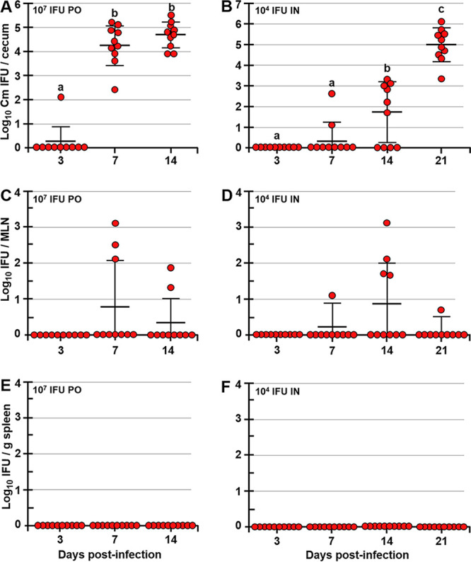
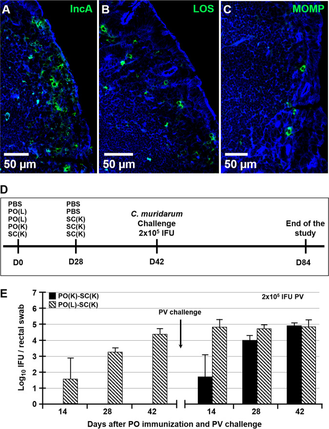
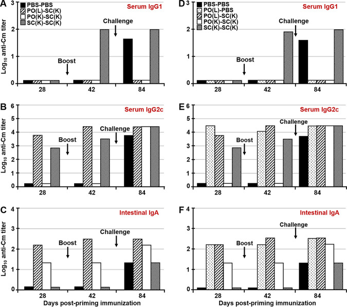
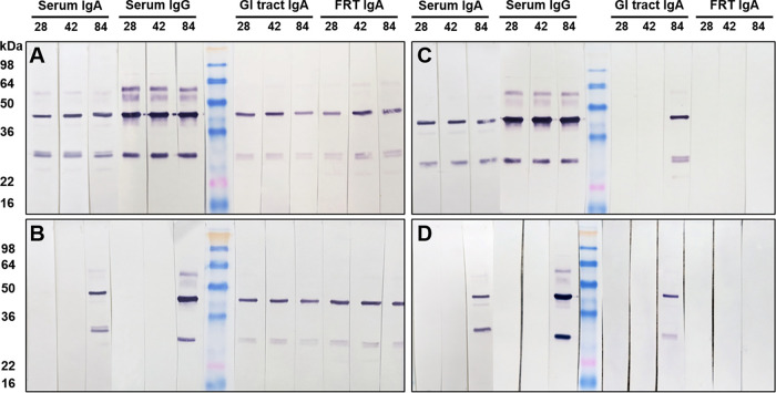
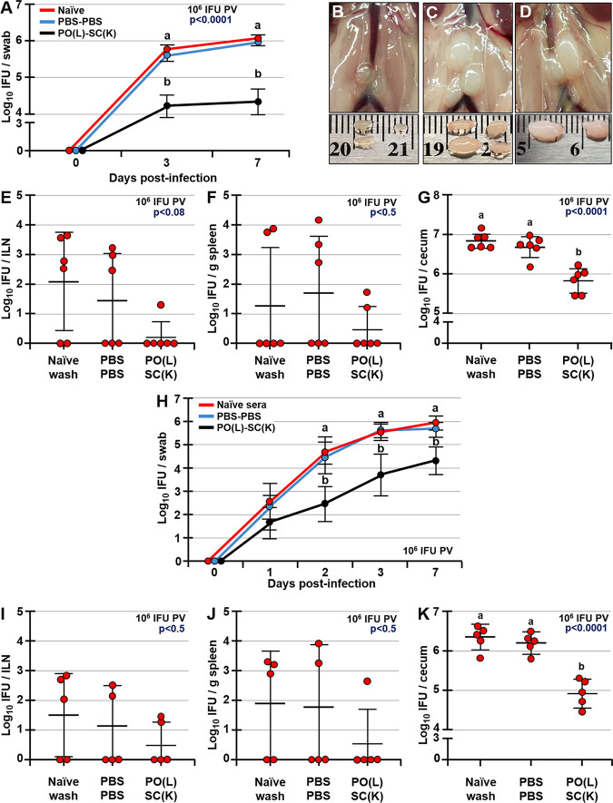
Chlamydia trachomatis is an obligate intracellular pathogen that causes sexually transmitted disease. In women, chlamydial infections may cause pelvic inflammatory disease (PID), ectopic pregnancy, and infertility. The role of antibodies in protection against a primary Chlamydia infection is unclear and was a focus of this work. Using the C. muridarum mouse infection model, we show that intestinal mucosa is infected via intranasal (i.n.) or per-oral (p.o.) Chlamydia inoculation and that unlike the female reproductive tract (FRT) mucosa, it halts systemic Chlamydia dissemination. Moreover, p.o. immunization or infection with Chlamydia confers protection against per-vaginal (p.v.) challenge, resulting in significantly decreased bacterial burden in the FRT, accelerated Chlamydia clearance, and reduced hydrosalpinx pathology. In contrast, subcutaneous (s.c.) immunization conferred no protection against the p.v. challenge. Both p.o. and s.c. immunizations induced Chlamydia-specific serum IgA. However, IgA was found only in the vaginal washes and fecal extracts of p.o.-immunized animals. Following a p.v. challenge, unimmunized control and s.c.-s.c.-immunized animals developed Chlamydia-specific intestinal IgA yet failed to develop IgA in the FRT, indicating that IgA response in the FRT relies on the FRT to gastrointestinal tract (GIT) antigen transport. Vaginal secretions of p.o.-immunized animals neutralize Chlamydia in vivo, resulting in significantly lower Chlamydia burden in the FRT and Chlamydia transport to the GIT. We also show that infection of the GIT is not necessary for induction of protective immunity in the FRT, a finding that is important for the development of p.o. subunit vaccines to target Chlamydia and possibly other sexually transmitted pathogens.

Keywords: Chlamydia; IgA; antibodies; female reproductive tract; mucosa; mucosal vaccines; neutralizing antibodies; vaccination; vaccine.


Figures

FIG 1
FIG 1
Following p.o. or i.n. administration, Chlamydia infects the GIT of mice but does not disseminate to the spleen. Shown are C. muridarum titers in ceca (A and B), MLNs (C and D), and spleens (E and F) of p.o.- or i.n.-infected mice. Mice were infected p.o. via gastric gavage with 107 or i.n. with 104 IFU of Chlamydia and at 3, 7, 14, and 21 dpi, and Chlamydia titers in tissues were determined. Chlamydia titers are expressed as log10 number of IFU per cecum, per MLN, or per gram of spleen. Data are representative of those from two separate experiments and are expressed as means ± SDs. Group means were separated using Tukey’s multiple-comparison test and were declared significantly different at a P value of <0.05 (n = 10 mice per time point). Group means that do not share a lowercase letter are significantly different.
FIG 2
FIG 2
Chlamydia inclusions in ceca of p.o.-infected mice and immunization and challenge timeline. Shown are chlamydial inclusions in ceca of mice p.o. infected with 107 IFU of C. muridarum at 7 dpi. Tissues were stained with antibodies specific for Chlamydia antigens IncA (A), LOS (B), or MOMP (C) (green) and actin-binding phalloidin-Alexa Fluor 350 (blue). (D) Timeline of prime-boost immunizations and p.v. challenge with C. muridarum. Mice were primed p.o. or s.c. with live or killed Chlamydia EBs at day 0 and then s.c. boosted at day 28 with killed EBs. Two weeks later (day 42), mice were p.v. challenged with 2 × 105 IFU of C. muridarum. Six weeks after p.v. challenge (day 84), the studies were terminated and FRT pathology and Chlamydia burdens were determined. (E) Rectal swab titers of PO(K)-SC(K) and PO(L)-SC(K) mice collected at days 14, 28, and 42 prior to and 14, 28, and 42 after p.v. challenge with 2 × 105 IFU of Chlamydia.
FIG 3
FIG 3
Serum and mucosal antibody titers following p.o. immunization or challenge with C. muridarum. Antibody titers in sera and intestinal extracts of mice 4 weeks after priming (day 28), 2 weeks after boosting (day 42), and 6 weeks after p.v. challenge (day 84). Two studies were conducted. In the first study, animals were fed live or killed Chlamydia EBs via a pipette, while in the second study (D to F), Chlamydia live or killed EBs were administered by gastric gavage, in order to preclude sublingual immunization. Samples from five animals from treatment group of each study were pooled and Chlamydia-specific IgG1, IgG2c, and intestinal IgA were analyzed by ELISA. Titers are expressed as log10 values of IgG1, IgG2c, or IgA antibodies detected in serum (A, B, D, and E) or fecal extracts (C and F) of mice following priming, boosting, and p.v. challenge with C. muridarum. Titers represent the highest dilutions of samples showing an absorbance value at 405 nm that is twice that of the negative control.
FIG 4
FIG 4
Immunization does not affect Chlamydia loads in ceca, but it enhances Chlamydia clearance from the FRT following a p.v. challenge. (A and B) Chlamydia titers in ceca of unimmunized and immunized mice at 6 weeks after p.v. challenge with 2 × 105 IFU of C. muridarum. (C and D) Chlamydia titers in vaginal swabs of mice at days 3, 7, 14, 21, 35, and 42 after p.v. challenge with 2 × 105 IFU of C. muridarum. Chlamydia titers are expressed as log10 number of IFU per cecum or per swab. Data are expressed as means ± SDs. Group means were separated using Tukey’s multiple-comparison procedures and were declared significantly different at a P value of <0.05. Means that do not share a lowercase letter are significantly different from each other.
FIG 5
FIG 5
p.o. priming with live or killed C. muridarum significantly reduces FRT pathology and Chlamydia loads in ovaries/oviducts and uteri of mice following a p.v. challenge. (A and D) Hydrosalpinx scores and Chlamydia loads in ovaries/oviducts (B and E) and uteri (C and F) of C57BL/6J mice at 6 weeks after p.v. challenge with 2 × 105 IFU of C. muridarum. Chlamydia titers are expressed as log10 number of IFU per ovary/oviduct or uterus. Data are expressed as means ± SDs. Group means were separated using Tukey’s multiple-comparison procedures and were declared significantly different at a P value of <0.05. Means that do not share a lowercase letter are significantly different from each other.
FIG 6
FIG 6
Serum from immunized and p.v. challenged animals neutralizes Chlamydia EBs in vitro. Neutralizing activity of sera from immunized and unimmunized control mice collected after priming (day 28) (A and D), boosting (day 42) (B and E) and p.v. challenge (C and F) was determined. Pooled serum samples were collected from the first (A to C) and second (D to F) studies. Group means were separated by Tukey’s multiple-comparison procedures. Data are shown as means ± SDs and were declared statistically significant at a P value of <0.05. Means that do not share a lowercase letter are significantly different from each other.
FIG 7
FIG 7
Chlamydia-specific serum, intestinal, and FRT antibodies at 4 weeks after priming (day 28), 2 weeks after boosting (day 42), and 6 weeks after p.v. challenge (day 84). For Western blot analysis, nitrocellulose membranes containing whole Chlamydia antigen were incubated with pooled serum or vaginal wash samples overnight. Serum or vaginal wash IgG or IgA antibodies bound to Chlamydia antigens on the membrane were detected with AP-conjugated rabbit anti-mouse IgG or IgA antibodies.
FIG 8
FIG 8
MOMP-specific antibodies in sera and vaginal washes of immunized and control mice 2 weeks after boosting (day 42) and 6 weeks after p.v. challenge (day 84). MOMP-specific serum IgG (A and B) and vaginal wash IgA (C and D) antibodies after boosting immunization (day 42) (A and C) and after p.v. challenge (day 84) (B and D) are shown. Membranes harboring MOMP antigen were incubated overnight with pooled sera or vaginal washes (collected from two studies), and then MOMP-specific IgG and IgA antibodies were detected with AP-conjugated rabbit anti-mouse IgG or IgA antibodies.
FIG 9
FIG 9
IgA antibodies in vaginal washes of PO-immunized mice neutralize C. muridarum EBs and reduce systemic Chlamydia spread. (A and H) Chlamydia titers in vaginal swabs of naive mice prior to p.v. infection (day 0) and at 1, 2, 3, and 7 dpi p.v. with 106 IFU of EBs that were neutralized with either vaginal wash of naive (unimmunized and unchallenged) mice or PBS-PBS (unimmunized and p.v. challenged) and PO(L)-SC(K) mice collected 6 weeks postchallenge (day 84). (B and D) Example images of ILNs of mice infected with EBs neutralized with vaginal washes of PO(L)-SC(K) (B), PBS-PBS (C), or naive (unimmunized and unchallenged) (D) mice collected 6 weeks post-p.v. challenge (day 84). (E to G and I to K) Chlamydia titers in ILNs, spleens, and ceca of naive mice at 7 dpi p.v. with neutralized EBs as described for panel A. Data are expressed as means ± SDs. Means were separated using Tukey’s multiple-comparison procedures and are declared significantly different at a P value of <0.05 (n = 6 and n = 5). Means that do not share a lowercase letter are significantly different from each other.

Similar articles

Cited by

References

    1. WHO. 2019. Sexually transmitted infections (STIs). https://www.who.int/en/news-room/fact-sheets/detail/sexually-transmitted.... Accessed 16 June 2019.
    1. CDC. 2018. Sexually transmitted disease surveillance 2018. https://www.cdc.gov/std/stats18/default.htm.
    1. Brunham RC, Gottlieb SL, Paavonen J. 2015. Pelvic inflammatory disease. N Engl J Med 372:2039–2048. doi:10.1056/NEJMra1411426. - DOI - PubMed
    1. Soper DE, Brockwell NJ, Dalton HP. 1992. Microbial etiology of urban emergency department acute salpingitis: treatment with ofloxacin. Am J Obstet Gynecol 167:653–660. doi:10.1016/s0002-9378(11)91566-x. - DOI - PubMed
    1. Haggerty CL, Gottlieb SL, Taylor BD, Low N, Xu F, Ness RB. 2010. Risk of sequelae after Chlamydia trachomatis genital infection in women. J Infect Dis 201(Suppl 2):S134–S155. doi:10.1086/652395. - DOI - PubMed

Publication types

MeSH terms