. 2020 Nov 2;192(44):E1383-E1386.
doi: 10.1503/cmaj.200641-f.

Prélèvements nasopharyngés initialement négatifs chez un homme de 76 ans infecté par le SRAS-CoV-2

[Article in French]
Affiliations

Prélèvements nasopharyngés initialement négatifs chez un homme de 76 ans infecté par le SRAS-CoV-2

[Article in French]
Divjot S Kumar et al. CMAJ. .
No abstract available


Conflict of interest statement

Intérêts concurrents: Aucun déclaré.

Figures
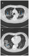
Figure 1:
Figure 1:
Clichés de TDM thoracique (A) et (B) (pris au jour 3 d’hospitalisation) chez un homme de 76 ans porteur de la maladie à coronavirus 2019 (COVID-19) et ayant eu des résultats négatifs aux prélèvements nasopharyngés. On observe une hyperdensité périphérique bilatérale en verre dépoli avec zones de lignes septales constituant une mosaïque (flèches bleues). Il s’agit d’un aspect typique de la COVID-19, selon l’énoncé consensuel des experts de la Radiological Society of North America.
Figure 2:
Figure 2:
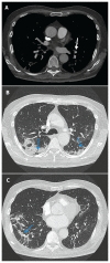
Clichés de TDM thoracique pris au jour 15 d’hospitalisation. (A) Embolie pulmonaire segmentaire non occlusive au lobe inférieur gauche (flèche blanche). (B) et (C) Amélioration de l’aspect en verre dépoli et épaississement septal; consolidation péribronchovasculaire et périlobulaire périphérique bilatérale avec légère perte de volume et distorsion parenchymateuse concordant avec l’évolution type d’une pneumonie organisée en cours d’évolution (flèches bleues).

References

    1. Simpson S, Kay FU, Abbara S, et al. Radiological Society of North America Expert Consensus Statement on Reporting Chest CT Findings Related to COVID-19. Endorsed by the Society of Thoracic Radiology, the American College of Radiology, and RSNA. Radiology: Cardiothoracic Imaging 2020. Mar. 25;2:e200152. 10.1148/ryct.2020200152. - DOI - PMC - PubMed
    1. Ai T, Yang Z, Hou H, et al. Correlation of chest CT and RT–PCR testing in coronavirus disease 2019 (COVID-19) in China: a report of 1014 cases. Radiology 2020. Feb. 26 [Cyberpublication avant impression]. 200642. 10.1148/radiol.2020200642. - DOI - PMC - PubMed
    1. Xie C, Jiang L, Huang G, et al. Comparison of different samples for 2019 novel coronavirus detection by nucleic acid amplification tests. Int J Infect Dis 2020; 93:264–7. - PMC - PubMed
    1. Winichakoon P, Chaiwarith R, Liwsrisakun C, et al. Negative nasopharyngeal and oropharyngeal swab does not rule put COVID-19. J Clin Microbiol 2020. Feb. 26 [Cyberpublication avant impression]. pii JCM.00297–20. 10.1128/JCM.00297-20. - DOI - PMC - PubMed
    1. Li W, Moore MJ, Vasilieva N, et al. Angiotensin-converting enzyme 2 is a functional receptor for the SARS coronavirus. Nature 2003;426:450–4. - PMC - PubMed