. 2020 Nov 18;12(570):eabd3876.
doi: 10.1126/scitranslmed.abd3876. Epub 2020 Nov 2.

Prothrombotic autoantibodies in serum from patients hospitalized with COVID-19

Affiliations

Prothrombotic autoantibodies in serum from patients hospitalized with COVID-19

Yu Zuo et al. Sci Transl Med. .

Abstract

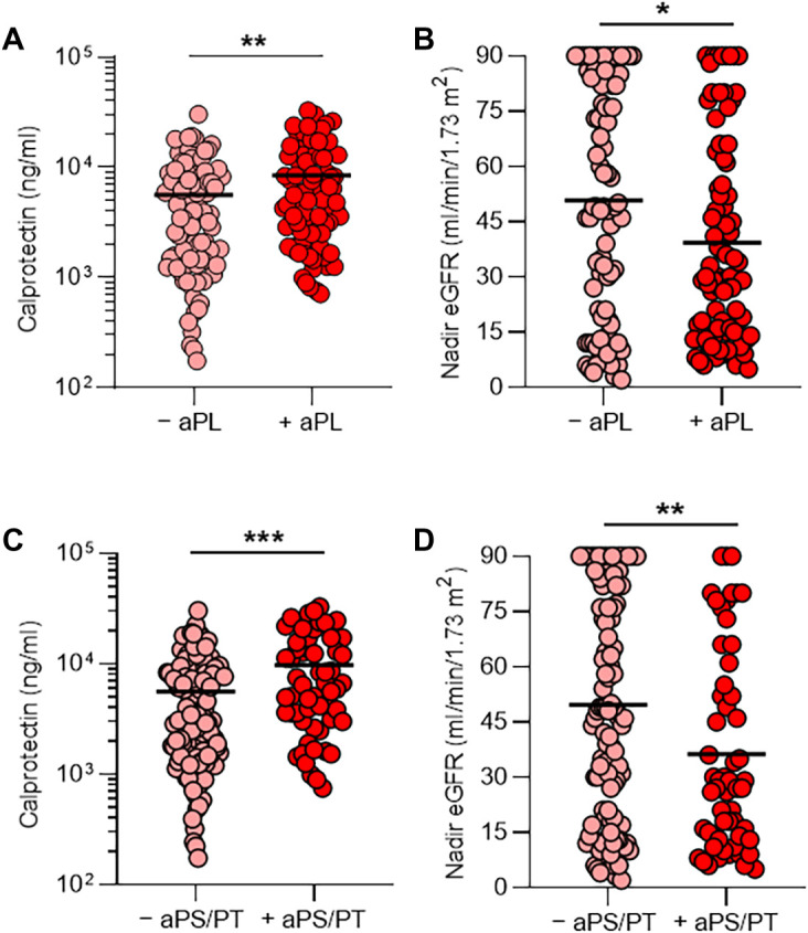
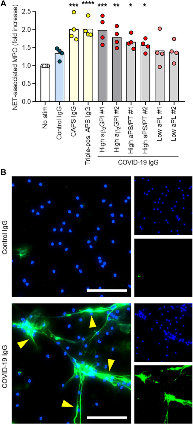
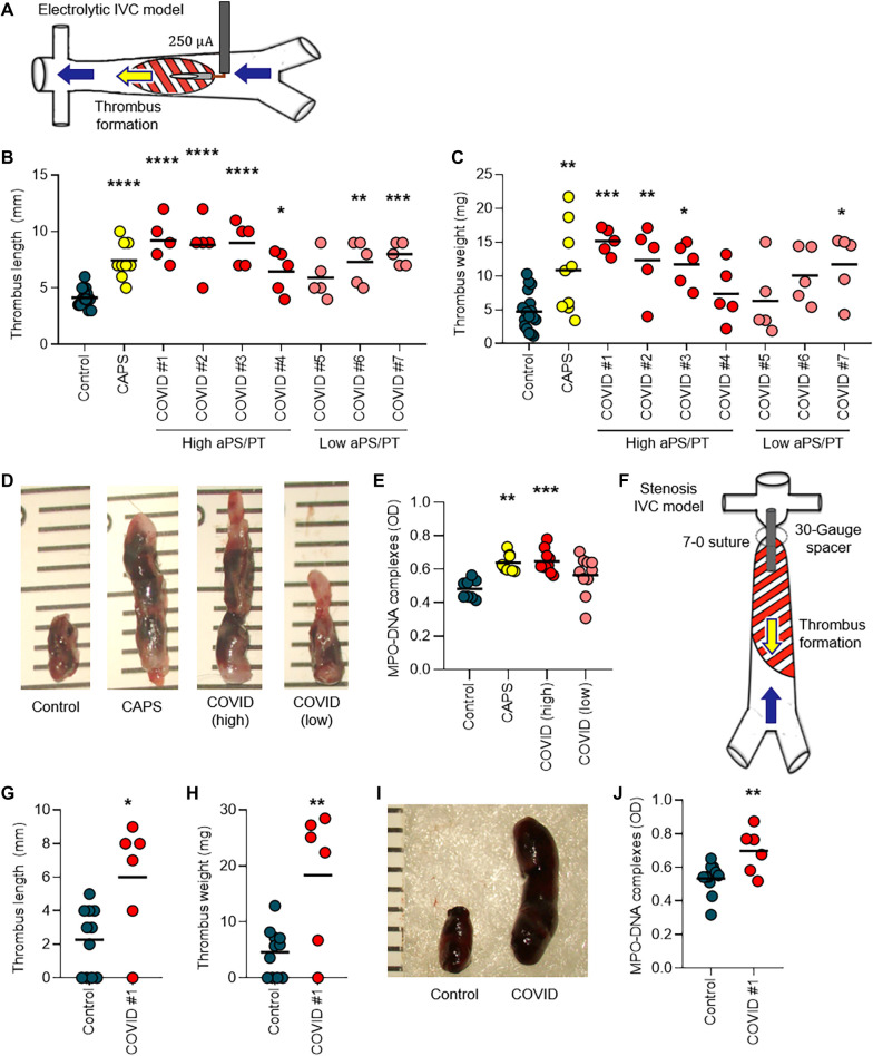
Patients with COVID-19 are at high risk for thrombotic arterial and venous occlusions. Lung histopathology often reveals fibrin-based blockages in the small blood vessels of patients who succumb to the disease. Antiphospholipid syndrome is an acquired and potentially life-threatening thrombophilia in which patients develop pathogenic autoantibodies targeting phospholipids and phospholipid-binding proteins (aPL antibodies). Case series have recently detected aPL antibodies in patients with COVID-19. Here, we measured eight types of aPL antibodies in serum samples from 172 patients hospitalized with COVID-19. These aPL antibodies included anticardiolipin IgG, IgM, and IgA; anti-β2 glycoprotein I IgG, IgM, and IgA; and anti-phosphatidylserine/prothrombin (aPS/PT) IgG and IgM. We detected aPS/PT IgG in 24% of serum samples, anticardiolipin IgM in 23% of samples, and aPS/PT IgM in 18% of samples. Antiphospholipid autoantibodies were present in 52% of serum samples using the manufacturer's threshold and in 30% using a more stringent cutoff (≥40 ELISA-specific units). Higher titers of aPL antibodies were associated with neutrophil hyperactivity, including the release of neutrophil extracellular traps (NETs), higher platelet counts, more severe respiratory disease, and lower clinical estimated glomerular filtration rate. Similar to IgG from patients with antiphospholipid syndrome, IgG fractions isolated from patients with COVID-19 promoted NET release from neutrophils isolated from healthy individuals. Furthermore, injection of IgG purified from COVID-19 patient serum into mice accelerated venous thrombosis in two mouse models. These findings suggest that half of patients hospitalized with COVID-19 become at least transiently positive for aPL antibodies and that these autoantibodies are potentially pathogenic.


Figures

Fig. 1
Fig. 1. aPL antibodies, NET release, and renal function.
Serum samples were obtained from 172 patients hospitalized with COVID-19. (A and B) Patients were divided into two groups on the basis of whether their serum samples were positive (+) or negative (−) for the presence of aPL antibodies (positivity was based on the manufacturer’s threshold). Shown is the amount of calprotectin in serum, a measure of neutrophil activation (A), and the clinical estimated glomerular filtration rate (eGFR) (B) for the two groups. (C and D) Patients were divided into two groups on the basis of whether their serum samples were positive (+) or negative (−) for the presence of aPS/PT antibodies (IgG and IgM considered together); the manufacturer’s thresholds were used to determine positivity. Shown is the amount of calprotectin (C) and the eGFR (D) for the two groups. Groups were analyzed by an unpaired t test: *P < 0.05, **P < 0.01, and ***P < 0.001. Horizontal black bars represent the mean. For patients who had serum samples available at multiple time points, only the first available serum sample was used in this analysis.
Fig. 2
Fig. 2. COVID-19 patient IgG promotes NET release from normal neutrophils in vitro.
(A) Control neutrophils were isolated from healthy individuals and cultured in the presence of human IgG (10 μg/ml) for 3 hours. IgG fractions were obtained from patients with COVID-19 who were or were not positive for aPL antibodies (aPS/PT or aβ2GPI as indicated), and from patients with antiphospholipid syndrome (APS) or catastrophic APS (CAPS). NET release was measured by the enzymatic activity of myeloperoxidase (MPO) after solubilization of NETs with micrococcal nuclease; fold increase is plotted relative to unstimulated neutrophils (no stim). Data are derived from four independent experiments. Comparisons were to the unstimulated group by one-way ANOVA with correction for multiple comparisons by Dunnett’s method: *P < 0.05, **P < 0.01, ***P < 0.001. (B) Representative images show released NETs, indicated by yellow arrows. DNA, blue; neutrophil elastase, green. Scale bars, 100 μm.
Fig. 3
Fig. 3. IgG from patients with COVID-19 potentiates thrombosis in mice.
(A) Schematic shows thrombus initiation in the inferior vena cava (IVC) of mice by local electrolysis leading to free radical generation and activation of the endothelium. (B and C) Mice were administered IgG from healthy individuals (control), from patients with COVID-19 who had high or low aPS/PT antibodies, or from patients with catastrophic APS (CAPS). Just before intravenous administration of IgG, mice were subjected to local electrolysis in the IVC. Thrombus length (B) and weight (C) were determined 24 hours after IgG injection. Scatter plots with individual data points (each point represents a single mouse) are presented. (D) Shown are photographs of representative thrombi from the experiments presented in (B) and (C). The rulers are measuring thrombi in millimeters. (E) Serum samples from mice in the experiments presented in (B) and (C) were tested for NET remnants measured by an ELISA that detected myeloperoxidase (MPO)–DNA complexes. Scatter plots with individual data points (each point represents a single mouse) are presented. OD, optical density. (F) Schematic shows thrombus initiation in the IVC of mice by a stenosis that was induced via placement of a fixed suture over a spacer that was subsequently removed. (G and H) Mice were treated intravenously with IgG from a healthy individual (control) or from a patient with COVID-19 with high aPS/PT antibodies. Just before intravenous administration of IgG, stenosis was induced. Twenty-four hours later, thrombus length (G) and weight (H) were determined. Scatter plots with individual data points (each point represents a single mouse) are presented. (I) Shown are photographs of representative thrombi from the experiments presented in (G) and (H). (J) Serum samples from mice in the experiments presented in (G) and (H) were tested for NET remnants measured by an ELISA that detected MPO-DNA complexes. Scatter plots with individual data points (each point represents a single mouse) are presented. Horizontal black bars represent the mean. Comparisons were by either one-way ANOVA with correction for multiple comparisons by Dunnett’s method (B, C, and E) or unpaired t test (G, H, and J): *P < 0.05, **P < 0.01, ***P < 0.001, and ****P < 0.0001.

Update of

Similar articles

Cited by

References

    1. Wu C., Chen X., Cai Y., Xia J., Zhou X., Xu S., Huang H., Zhang L., Zhou X., Du C., Zhang Y., Song J., Wang S., Chao Y., Yang Z., Xu J., Zhou X., Chen D., Xiong W., Xu L., Zhou F., Jiang J., Bai C., Zheng J., Song Y., Risk factors associated with acute respiratory distress syndrome and death in patients with coronavirus disease 2019 pneumonia in Wuhan, China. JAMA Intern. Med. 180, 934–943 (2020). - PMC - PubMed
    1. Tang N., Li D., Wang X., Sun Z., Abnormal coagulation parameters are associated with poor prognosis in patients with novel coronavirus pneumonia. J. Thromb. Haemost. 18, 844–847 (2020). - PMC - PubMed
    1. Guan W.-j., Ni Z.-y., Hu Y., Liang W.-h., Ou C.-q., He J.-x., Liu L., Shan H., Lei C.-l., Hui D. S. C., Du B., Li L.-j., Zeng G., Yuen K.-Y., Chen R.-c., Tang C.-l., Wang T., Chen P.-y., Xiang J., Li S.-y., Wang J.-l., Liang Z.-j., Peng Y.-x., Wei L., Liu Y., Hu Y.-h., Peng P., Wang J.-m., Liu J.-y., Chen Z., Li G., Zheng Z.-j., Qiu S.-q., Luo J., Ye C.-j., Zhu S.-y., Zhong N.-s.; China Medical Treatment Expert Group for Covid-19 , Clinical characteristics of coronavirus disease 2019 in China. N. Engl. J. Med. 382, 1708–1720 (2020). - PMC - PubMed
    1. Chen N., Zhou M., Dong X., Qu J., Gong F., Han Y., Qiu Y., Wang J., Liu Y., Wei Y., Xia J., Yu T., Zhang X., Zhang L., Epidemiological and clinical characteristics of 99 cases of 2019 novel coronavirus pneumonia in Wuhan, China: A descriptive study. Lancet 395, 507–513 (2020). - PMC - PubMed
    1. Zhou F., Yu T., Du R., Fan G., Liu Y., Liu Z., Xiang J., Wang Y., Song B., Gu X., Guan L., Wei Y., Li H., Wu X., Xu J., Tu S., Zhang Y., Chen H., Cao B., Clinical course and risk factors for mortality of adult inpatients with COVID-19 in Wuhan, China: A retrospective cohort study. Lancet 395, 1054–1062 (2020). - PMC - PubMed

Publication types

MeSH terms

Substances