. 2020 Nov 17;117(46):29155-29165.
doi: 10.1073/pnas.2016539117. Epub 2020 Nov 2.

LRRC8 family proteins within lysosomes regulate cellular osmoregulation and enhance cell survival to multiple physiological stresses

Affiliations

LRRC8 family proteins within lysosomes regulate cellular osmoregulation and enhance cell survival to multiple physiological stresses

Ping Li et al. Proc Natl Acad Sci U S A. .

Abstract

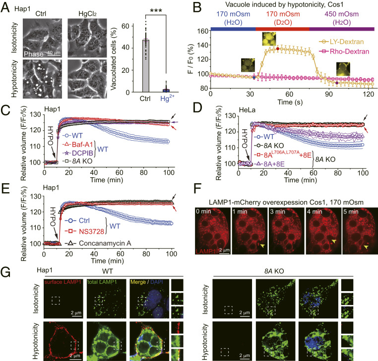
LRRC8 family proteins on the plasma membrane play a critical role in cellular osmoregulation by forming volume-regulated anion channels (VRACs) necessary to prevent necrotic cell death. We demonstrate that intracellular LRRC8 proteins acting within lysosomes also play an essential role in cellular osmoregulation. LRRC8 proteins on lysosome membranes generate large lysosomal volume-regulated anion channel (Lyso-VRAC) currents in response to low cytoplasmic ionic strength conditions. When a double-leucine L706L707 motif at the C terminus of LRRC8A was mutated to alanines, normal plasma membrane VRAC currents were still observed, but Lyso-VRAC currents were absent. We used this targeting mutant, as well as pharmacological tools, to demonstrate that Lyso-VRAC currents are necessary for the formation of large lysosome-derived vacuoles, which store and then expel excess water to maintain cytosolic water homeostasis. Thus, Lyso-VRACs allow lysosomes of mammalian cells to act as the cell`s "bladder." When Lyso-VRAC current was selectively eliminated, the extent of necrotic cell death to sustained stress was greatly increased, not only in response to hypoosmotic stress, but also to hypoxic and hypothermic stresses. Thus Lyso-VRACs play an essential role in enabling cells to mount successful homeostatic responses to multiple stressors.

Keywords: chloride channel; exocytosis; lysosome; osmoregulation; vacuolation.


Conflict of interest statement

The authors declare no competing interest.

Figures

Fig. 1.
Fig. 1.
Hypotonicity induces lysosome vacuolation and induces a chloride current in vacuoles. (A) Responses of Cos1, Hap1, and HeLa cells to a hypotonic challenge. Thirty minutes prior to data collection, the complete medium was replaced with fresh complete medium (Isotonicity, 300 mOsm) or the test medium (Hypotonicity, 170 mOsm). The photomicrographs show phase contrast images with examples of the vacuoles (bright white spots) commonly observed in cells exposed to hypotonic solutions. Also see Movie S1. The Lower quantifies data from many similar images with the parameter “Vacuolated Cell (%),” indicating the proportion of cells with at least one vacuole. In all panels with asterisks * is P < 0.05. ** is P < 0.01. *** is P < 0.001. (B) Each group of images shows cells studied after 0.5 h in either 300 mOsm (Upper) or 170 mOsm (Lower) medium. Each panel shows superimposed DIC (differential interference contrast) and fluorescence images. The red signal is LAMP1-mCherry driven by transient transfection, and the green signal is Dextran-Green loaded into the cell by endocytosis. The graph to the Right of each panel shows the fluorescence intensity of a line scan (blue line on the blown-up image) through the double labeled object indicated by the white arrow. (C) Images demonstrating the whole-endolysosome (whole-EL) patch-clamp configuration being achieved on a vacuolated lysosome that had been induced by hypotonic (80 mOsm) challenge. Note that approximately half the cell had been torn away to make the vacuole accessible for recording. See also SI Appendix, Fig. S2A. (D and E) Representative currents on vacuoles of Cos1 cells induced by exposure to 80 mOsm solution. The difference between the currents in 320 mOsm cytosolic solution and 80 mOsm cytosolic solution is defined as Lyso-VRAC. The currents in D were elicited by 200-ms ramps from −120 mV to +120 mV with the membrane potential (Vm) at each time point indicated on the x axis. The currents in E were elicited by voltage steps using the protocol shown to the right of the current traces. The dashed line indicates 0 pA. (F) The time course of cytosolic ionic strength effects on Lyso-VRAC currents at ±120 mV obtained from traces like those illustrated in G. (G) Representative I-V (current-voltage) traces from vacuoles of Cos1 cells treated with vacuolin in response to various cytosol-side solutions (Basal, 140 mM K-Gluconate, 290 mOsm; Γi 40, 40 mM CsCl, 290 mOsm; Γi 140, 140 mM CsCl, 290 mOsm). (H) Intracellular ionic strength (Γi) dependence of Lyso-VRAC. (I) Reversal potential of the Lyso-VRAC current was dependent on [Cl]Lumen. The slope of the line fit to the data was −46.1 ± 0.5 mV per 10-fold concentration change. (J) ILyso-VRAC induced by Γi 40 in additional cell types. In addition to Cos1, HeLa, and Hap1 cells, the cell types shown are Hap1 cells in which the endogenous LRRC8A gene was replaced with a sequence encoding an LRRC8A-GFP fusion protein (GFP-KI Hap1), HEK293T, human breast cancer cells (MDA231), mouse embryonic fibroblasts (MEF), and WT mouse bone marrow macrophages (BMM). For all panels showing averaged data, n > 6 and Student’s t tests were used to determine statistical significance. pA, picoamperes; pF, picofarads.
Fig. 2.
Fig. 2.
LRRC8 proteins are required to produce Lyso-VRAC. (A) ILyso-VRAC from endolysosomes enlarged with vacuolin-1 in WT and 8A KO Hap1 cells with basal and low ionic strength bath solutions. [Cl]Pipette/Lumen = 140mM, and [Cl] = 40mM for Γi 40. DCPIB was applied in the Γi 40 solution. (B) ILyso-VRAC in 8A KO Hap1 cells dually transfected with LRRC8A-mCherry and LRRC8E-GFP. (C) Current density of ILyso-VRAC in WT and in 8A KO Hap1 with or without overexpressed LRRC8 proteins. Current densities at ±120 mV are shown. Data for each condition were assessed for significance using Student`s t test and were collected from six or more independent experiments. *** is P < 0.001 and “N.S.” is P > 0.05. (D) Subcellular fractionation analysis of LRRC8A-GFP expression in various organelle fractions of LRRC8A-GFP–knock-in Hap1 cells. The organelle-specific markers identified in these Western blots were as follows: Lysosomes, LAMP1 and Cathepsin D; Mitochondria, Complex II; Early Endosomes, EEA1; and the Golgi apparatus, GM130. Similar data were collected from three independent experiments, and the graph shows the means ± SEMs. (E) The expression pattern of LRRC8A-GFP when this coding sequence was knocked into the endogenous location of LRRC8A in Hap1 cells. Lysosomes were labeled by immunostaining for LAMP1 (red). The boxed regions in the Upper images are shown at higher magnification in the Lower images. The graph below each group of images is a line scan through the plasma membrane and lysosome shown in the boxed region indicating the intensity of 8A-GFP (green lines) and Anti-LAMP1 (red lines) along the blue line. (F) Images and line scan analysis showing the colocalization of 8A-mCherry (red), 8E-BFP (blue), and LAMP1-GFP (green) in transfected Cos1 cells.
Fig. 3.
Fig. 3.
A double-Leucine motif is required for lysosomal localization of LRRC8A to mediate Lyso-VRAC. (A) Images and line scan analysis showing the lack of colocalization of 8AL706A,L707A-mCherry (red) and LAMP1-GFP (green) in transfected Cos1 cells. See SI Appendix, Fig. S5G for evidence that the 8AL706A,L707A-mCherry staining is in the ER. (B) Images and line scan analysis showing that 8AL706A,L707A-mCherry (red) and 8E-BFP (blue) localized to a different organelle than LAMP1-GFP (green) in transfected Cos1 cells. (C) Representative traces of ILyso-VRAC in 8A KO Hap1 cells dually transfected with 8AL706A,L707A-mCherry and 8E-GFP. (D) Current density of ILyso-VRAC in WT and in 8A KO Hap1 with or without overexpressed 8AL706A,L707A-mCherry and 8E-GFP. Data for each condition in this panel and in I were assessed for significance using Student`s t test and were collected from six or more independent experiments. (E and F) Whole cell recordings to test for plasma membrane VRAC (PM-VRAC) currents in WT and 8A KO Hap1 cells. These experiments used Γi 40 ([Cl] = 40 mM) as the cytosolic/pipette solution and Γi 140 ([Cl] = 140 mM) as the extracellular/bath solution. The responses are shown at 0 (red) and 400 s (black) after break-in. (G) Experiment similar to E except that the 8A KO Hap1 cells were cotransfected with 8A-mCherry and 8E-GFP. (H) Experiment similar to E except that the 8A KO Hap1 cells were transfected with 8AL706A,L707A-mCherry and 8E-GFP. (I) Current density of IPM-VRAC in WT and 8A KO Hap1 cells with or without LRRC8 proteins overexpressed. *** is P < 0.001 and “N.S.” is P > 0.05.
Fig. 4.
Fig. 4.
Lyso-VRAC is required for hypotonicity-induced lysosomal vacuolation. (A) Hypotonicity-induced cytoplasmic vacuolation in WT, 8A KO, TPC1&2 double KO (DKO), TMEM175 KO, and CLC7 KO Hap1 cells. Quantitative analysis is to the Right. In all panels with asterisks *** is P < 0.001 and N.S. is P > 0.05. (B and C) Sample images (Left) and quantitative analysis (Right) of lysosomal volume expansion (B) and lysosome vacuolation (C) under hypotonic stress in 8A KO HeLa cells that were dually transfected with 8E-BFP + 8A-mCherry or 8AL706A,L707A-mCherry. (D) Differential effects of NS3728 (30 µM; membrane-impermeable) and DCPIB (100 µM, membrane-permeable) VRAC inhibitors on hypotonicity-induced vacuolation in WT and 8A KO Hap1 cells. (E) Effect of 100 µM DCPIB and LRRC8A knock out on lysosomal volume. In all bar graphs, data points were collected from more than six independent experiments, and Student`s t test was used to determine significance.
Fig. 5.
Fig. 5.
Lysosomes store and expel intracellular water to regulate cytosolic RVD. (A) Representative images (Left) and quantitation (Right) of effects of 50 µM HgCl2 (an aquaporin inhibitor) on lysosome vacuolation induced by hypotonicity. *** is P < 0.001. (B) Fluorescence of Lucifer yellow dextran (LY-Dx, yellow) and Rhodamine dextran (Rho-Dx, red) in response to D2O- or H2O-containing hypotonic (170 mOsm) solutions in Cos1 cells. Also see Movie S3. LY-Dextran and Rho-Dextran were preloaded into lysosomes of Cos1 cells by endocytosis. (CE) Time course of regulatory cytosol volume decrease (RVD) in response to 110-mOsm solutions estimated by measuring the fluorescence intensity of Calcein-AM (41). The concentrations of drugs used were 5 µM Baf-A1, 100 µM DCPIB, 30 µM NS3728, and 20 µM Concanamycin A. (F) Snapshots of a Cos1 cell transfected with LAMP1-mCherry from a time-lapse series. Time 0 is defined as 78 min after the onset of the cell being placed in 170 mOsm medium to induce production of large vacuoles (also see Movie S4). Exocytosis events are indicated with yellow arrowheads at 1, 4, and 5 min. (G) Effects of hypotonic treatment (0.5 h) on LAMP1 surface staining in WT (Left) and 8A KO (Right) Hap1 cells. Cells were immunostained using an anti-LAMP1 antibody that recognized an extracellular/luminal epitope under conditions that first stained only plasma membrane LAMP1 (nonpermeabilized cells, red) and then total LAMP1 (permeabilized cells, green). Nuclei were counterstained with DAPI (blue). Zoomed-in images of the boxed regions are shown in Right. In BE, data points were collected from six independent experiments.
Fig. 6.
Fig. 6.
Lyso-VRAC protects cells from necrotic death to multiple stressors. Effects of 1 h hour of treatment with hypotonicity (80 mOsm; A and B), hypoxia (5% O2; C and D), and hypothermia (4 °C; E and F) on cell survival of Hap1 or HeLa cells. In all panels with asterisks *** is P < 0.001 and N.S. is P > 0.05. (A, C, and E) Representative images of cells stained with propidium iodide (red) to identify necrotic cells, viewed with phase contest microscopy. Cells with vacuoles are indicated with green arrowheads. (B, D, and F) Cell survival assayed by measuring extracellular LDH activity for samples from the indicated cell type. LDH activity for each group of cells was normalized as the percentage of the LDH activity released from the same number of cells by treatment with distilled water (which caused all cells to die). The survival index was then calculated as 100%-relative LDH activity.
Fig. 7.
Fig. 7.
Working model for how Lyso-VRAC–mediated lysosome osmoregulation behavior is protective for cell survival. Hypotonicity results in water influx through aquaporin channels on the plasma membrane (cyan), and the cell volume increases almost instantly (the yellow curve on the top). This decreases cytosolic ionic strength, normally activating VRAC channels (green) formed from LRRC8 proteins on both the plasma membrane (PM-VRAC) and in lysosomes (Lyso-VRAC). The activation of Lyso-VRAC (but not PM-VRAC) triggers lysosomal vacuolation (the purple curve on the top) through lysosomal osmotic swelling and/or homotypic fusion, creating large-volume compartments for intracellular water storage. Lysosome vacuolation decreases the cytosol volume of cells, helping to maintain the physiological concentrations of proteins and metabolites. Exocytosis of water-filled vacuoles substantially reduces the cell volume by dumping excess water into the extracellular environment and reduces plasma membrane tension stress by delivering more intracellular lipids to the cell surface (Survival, upper scenario). When Lyso-VRAC is absent, giant vacuoles do not form, the plasma membrane breaks, and cells die, even if PM-VRAC currents are normal (Death, lower scenario).

Similar articles

Cited by

References

    1. Delpire E., Gagnon K. B., Water homeostasis and cell volume maintenance and regulation. Curr. Top. Membr. 81, 3–52 (2018). - PMC - PubMed
    1. Hoffmann E. K., Lambert I. H., Pedersen S. F., Physiology of cell volume regulation in vertebrates. Physiol. Rev. 89, 193–277 (2009). - PubMed
    1. Bourque C. W., Central mechanisms of osmosensation and systemic osmoregulation. Nat. Rev. Neurosci. 9, 519–531 (2008). - PubMed
    1. Kültz D., Cellular osmoregulation: Beyond ion transport and cell volume. Zoology (Jena) 104, 198–208 (2001). - PubMed
    1. Jentsch T. J., VRACs and other ion channels and transporters in the regulation of cell volume and beyond. Nat. Rev. Mol. Cell Biol. 17, 293–307 (2016). - PubMed

Publication types