. 2020 Nov 17;117(46):29055-29062.
doi: 10.1073/pnas.2012480117. Epub 2020 Nov 2.

CEACAMs serve as toxin-stimulated receptors for enterotoxigenic Escherichia coli

Affiliations

CEACAMs serve as toxin-stimulated receptors for enterotoxigenic Escherichia coli

Alaullah Sheikh et al. Proc Natl Acad Sci U S A. .

Abstract

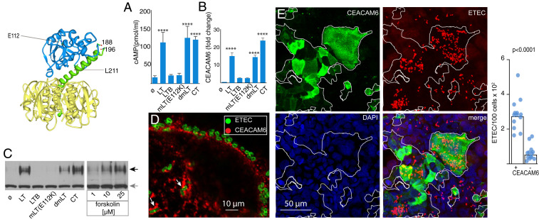
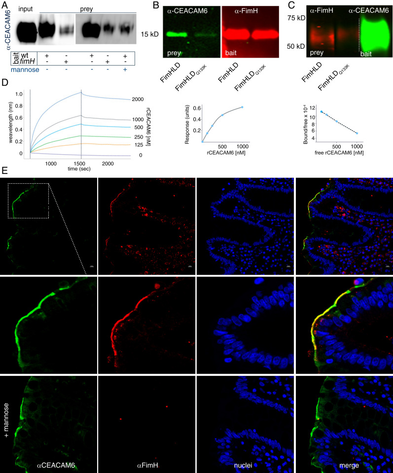
The enterotoxigenic Escherichia coli (ETEC) are among the most common causes of diarrheal illness and death due to diarrhea among young children in low-/middle-income countries (LMICs). ETEC have also been associated with important sequelae including malnutrition and stunting, placing children at further risk of death from diarrhea and other infections. Our understanding of the molecular pathogenesis of acute diarrheal disease as well as the sequelae linked to ETEC are still evolving. It has long been known that ETEC heat-labile toxin (LT) activates production of cAMP in the cell, signaling the modulation of cellular ion channels that results in a net efflux of salt and water into the intestinal lumen, culminating in watery diarrhea. However, as LT also promotes ETEC adhesion to intestinal epithelial cells, we postulated that increases in cAMP, a critical cellular "second messenger," may be linked to changes in cellular architecture that favor pathogen-host interactions. Indeed, here we show that ETEC use LT to up-regulate carcinoembryonic antigenrelated cell adhesion molecules (CEACAMs) on the surface of small intestinal epithelia, where they serve as critical bacterial receptors. Moreover, we show that bacteria are specifically recruited to areas of CEACAM expression, in particular CEACAM6, and that deletion of this CEACAM abrogates both bacterial adhesion and toxin delivery. Collectively, these results provide a paradigm for the molecular pathogenesis of ETEC in which the bacteria use toxin to drive up-regulation of cellular targets that enhances subsequent pathogen-host interactions.

Keywords: CEACAM; diarrhea; enterotoxin.


Conflict of interest statement

The authors declare no competing interest.

Figures

Fig. 1.
Fig. 1.
Heat-labile toxin stimulates production of CEACAM6 in intestinal epithelia. (Upper Left) Schematic depicts the heat-labile toxin (LT) with the A1, A2, and B subunits depicted in blue, green, and yellow, respectively, with the location of sites mutated in mLT (E112K) and dmLT (R192G, L211A). (A) cAMP activation in target Caco-2 epithelial cells following treatment with LT, LT mutants, the B subunit of LT alone (LTB), or cholera toxin (CT). ø, control baseline untreated cells (****P < 0.0001 by ANOVA comparison to untreated cells). (B) RT-PCR data demonstrating fold change in CEACAM6 expression relative to untreated cells normalized to GAPDH (mean ± SD, n = 3 technical replicates) relative to control (****P < 0.0001 by ANOVA). (C) Immunoblot of CEACAM6 expression following treatment with toxins or forskolin (Right). Black arrow indicates migration of CEACAM6, while gray arrow indicates migration of tubulin loading control. (D) Accumulation of CEACAM6 (red) adjacent to sites of ETEC (green) attachment to Caco-2 cells. (E) Confocal microscopic images showing association of ETEC with areas of CEACAM6 expression on Caco-2 cells. (Right) Bars on the graph indicate geometric means (****P < 0.0001 by Mann–Whitney U two-tailed nonparametric comparison of n = 13 replicates in each group).
Fig. 2.
Fig. 2.
CEACAM6 facilitates ETEC pathogen–host interactions. (A) CRISPR-Cas9–generated CEACAM6−/− mutant cell line (knockout, ko) does not make CEACAM6 (anti-CEACAM6 immunoblot; Left), but makes CEACAM1 and CEACAM5 (Right). Gray arrows indicate tubulin as a loading control. ø, untreated cells; LT and CT, stimulation with heat-labile or cholera toxin overnight, respectively. (B) Confocal images of wild type H10407 ETEC adherent to CEACAM6+/+ cells (Top) and CEACAM6-deficient cells (−/−; Bottom). (C) CEACAM6 is required for optimal interaction of ETEC with intestinal epithelial cells. (D) CEACAM6 is required for efficient toxin delivery by ETEC. Intracellular cAMP concentrations were determined in CEACAM6+/+ and −/− cells following infection with either wild type (wt) or control LT-negative eltA mutant strain (n = 15 replicates from a total of three independent experiments). (Inset) cAMP responses (in pmol/mL) of CEACAM6+/+ and −/− cells following treatment with forskolin. Symbols represent n = 5 replicates (bars represent geometric mean values; ****P < 0.0001 by Mann–Whitney U two-tailed nonparametric testing). (E and F) Restoration of CEACAM6 expression (red) in CEACAM6−/− mutant cell line G2 restores ETEC (green) adhesion. Graphs depict ETEC adhesion to G2 cells (−/+CEACAM6) expressed as bacteria per field 30 and 90 min after infection. (GI) Introduction of CEACAM6 into HeLa cells promotes ETEC adhesion.
Fig. 3.
Fig. 3.
CEACAM6 interacts with the FimH tip adhesin of type 1 fimbriae. (A) FimH promotes interaction of CEACAM6 with ETEC. Anti-CEACAM6 immunoblot shows amount of CEACAM6 retained by wild type (wt) and fimH mutant ETEC following incubation of bacteria with recombinant CEACAM6 (rCEACAM6). (B, Left) CEACAM6 far Western blot (green) in which either FimHLD or the FimHLDQ133K mutant protein was used as bait. (Right) Immunoblot (red) shows relative amounts of protein transferred to the blot as bait. (C, Left) FimH far Western blot in which immobilized CEACAM6 is used as bait for FimH or the FimHLDQ133K mutant. (Right) Transferred rCEACAM6 used as bait is shown. (D) Biolayer interferometry studies demonstrate interaction between immobilized rFimH and varying concentrations of rCEACAM6. (E) rFimHLD colocalizes with CEACAM6 on human small intestinal biopsies in a mannose-dependent fashion.
Fig. 4.
Fig. 4.
ETEC–CEACAM interactions on human small intestinal epithelia. (A) CEACAM6 is expressed on the apical surface of small intestinal enteroids. Arrows point to immunogold labeling of CEACAM6 on tips of microvilli (7,500×). (B) CEACAM6 associated with glycocalyx at tips of microvilli (15,000×). (C) ETEC (red) associates with CEACAM6 (green) on the surface of human small intestinal (Hu135D) epithelial cells (enteroids). Image prepared in Volocity from confocal image z-stacks. (D) Confocal microscopy images of “footprinting” of CEACAM6 (green) at sites of bacterial attachment (red) to small intestinal epithelia. Each panel is flanked by representations of the xz and yz planes (Bottom and Right, respectively). (E) ETEC induction of cAMP in polarized enteroid small intestinal monolayers. Shown are data for blood A (135D), B (248D), and O (235D). ø, untreated; PDEi, phosphodiesterase inhibition. (F) LT mediates up-regulation of CEACAM6 in small intestinal epithelia. Shown are experimental replicate data (n = 3) from control cells or cells treated with LT. (Inset) Immunoblot compares CEACAM6 protein expression in treated and untreated cells. (G) Heat-labile toxin promotes adhesion to small intestinal enteroids (shown are three independent experimental replicates, each with five to seven technical replicates, using enteroids derived from a donor belonging to a different ABO blood group). P values reflect nonparametric comparisons by Mann–Whitney U test. (H) CEACAM6 is required for optimal adhesion to small intestinal enteroids [**P = 0.0067 by ANOVA (Kruskal–Wallis) comparison of untreated (ø) or treatment with scramble RNA (sc) and CEACAM6 siRNA]. (Inset) Immunoblot demonstrates CEACAM6 production (top band) in enteroids by the ø, sc, si groups relative to tubulin (bottom band). (I) Human small intestinal biopsies demonstrating CEACAM6 expression on day 2, day 7, and day 30 post infection with ETEC. Control specimen is from the same patient (at day 7) processed without primary anti-CEACAM6 antibody.

References

    1. Khalil I. A., et al. , Morbidity and mortality due to shigella and enterotoxigenic Escherichia coli diarrhoea: The global burden of disease study 1990-2016. Lancet Infect. Dis. 18, 1229–1240 (2018). - PMC - PubMed
    1. Walker C. L. F., et al. , Global burden of childhood pneumonia and diarrhoea. Lancet 381, 1405–1416 (2013). - PMC - PubMed
    1. MAL-ED Network Investigators , The MAL-ED study: A multinational and multidisciplinary approach to understand the relationship between enteric pathogens, malnutrition, gut physiology, physical growth, cognitive development, and immune responses in infants and children up to 2 years of age in resource-poor environments. Clin. Infect. Dis. 59 (suppl. 4), S193–S206 (2014). - PubMed
    1. Sack R. B., The discovery of cholera–Like enterotoxins produced by Escherichia coli causing secretory diarrhoea in humans. Indian J. Med. Res. 133, 171–180 (2011). - PMC - PubMed
    1. Carpenter C. C., et al. , Clinical and physiological observations during an epidemic outbreak of non-vibrio cholera-like disease in Calcutta. Bull. World Health Organ. 33, 665–671 (1965). - PMC - PubMed

Publication types

MeSH terms