. 2020 Nov 17;117(46):29113-29122.
doi: 10.1073/pnas.2006700117. Epub 2020 Nov 2.

An evolutionarily acquired microRNA shapes development of mammalian cortical projections

Affiliations

An evolutionarily acquired microRNA shapes development of mammalian cortical projections

Jessica L Diaz et al. Proc Natl Acad Sci U S A. .

Abstract

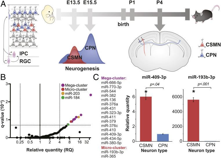
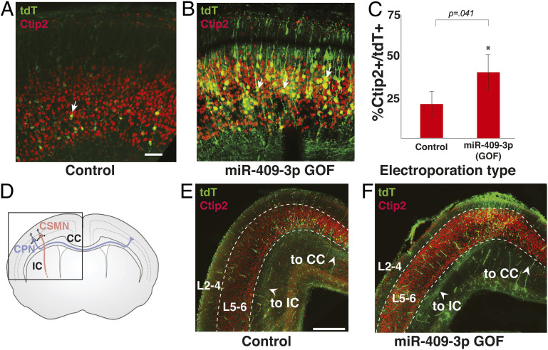
The corticospinal tract is unique to mammals and the corpus callosum is unique to placental mammals (eutherians). The emergence of these structures is thought to underpin the evolutionary acquisition of complex motor and cognitive skills. Corticospinal motor neurons (CSMN) and callosal projection neurons (CPN) are the archetypal projection neurons of the corticospinal tract and corpus callosum, respectively. Although a number of conserved transcriptional regulators of CSMN and CPN development have been identified in vertebrates, none are unique to mammals and most are coexpressed across multiple projection neuron subtypes. Here, we discover 17 CSMN-enriched microRNAs (miRNAs), 15 of which map to a single genomic cluster that is exclusive to eutherians. One of these, miR-409-3p, promotes CSMN subtype identity in part via repression of LMO4, a key transcriptional regulator of CPN development. In vivo, miR-409-3p is sufficient to convert deep-layer CPN into CSMN. This is a demonstration of an evolutionarily acquired miRNA in eutherians that refines cortical projection neuron subtype development. Our findings implicate miRNAs in the eutherians' increase in neuronal subtype and projection diversity, the anatomic underpinnings of their complex behavior.

Keywords: cerebral cortex; cortical development; microRNA; motor neuron; projection neuron.


Conflict of interest statement

Competing interest statement: C.J.S. and authors J.L.D., V.B.S., V.L., N.G.-N., M.B.W., R.N., Z.H., P.S., T.D.P., and S.T. are affiliated with Stanford University.

Figures

Fig. 1.
Fig. 1.
miRNAs are differentially expressed by CSMN vs. CPN during their development. (A) Schematic of CSMN (red) and CPN (blue) development in mice. IPC: Intermediate Progenitor Cell; RGC: Radial Glial Cell. (B) Volcano plot of differential miRNA expression by CSMN vs. CPN on P1, with fold change expressed as RQ plotted against statistical significance expressed as FDR-adjusted P value (q-value), reveals 19 miRNA candidates enriched at least 4-fold in CSMN relative to CPN with a q-value of less than 0.2 (colored dots). (C) Two confirmed miRNA clusters, comprising 17 differentially expressed candidate miRNAs, and representative results of independent validation via qPCR on P2. miR-409-3p from megacluster is 6-fold enriched (RQ = 6) in CSMN vs. CPN on P2; miR193b-3p is 5,630-fold enriched in CSMN vs. CPN on P2. Error bars represent SEM.
Fig. 2.
Fig. 2.
miR-409-3p is enriched in CSMN, and it represses the CPN-expressed and CSMN-excluded transcriptional regulator LMO4. (A) miR-409-3p is sixfold enriched in CSMN vs. CPN at P2 by qPCR. Error bars represent SEM. (B) LMO4 is enriched in CPN vs. CSMN in late embryonic and early postnatal life by microarray analysis. Error bars represent SEM. (C) Sequence alignments demonstrate that miR-409-3p is predicted to target two sites in the LMO4 3′ UTR. Seed sequence base pairing is shown in red. (D) miR-409-3p oligonucleotides repress a LMO4 3′ UTR luciferase reporter gene bearing wild-type, but not mismatch, miR-409-3p target sequences. Scrambled miRNA does not repress the LMO4 3′ UTR luciferase reporter. Error bars represent SEM. *P < 0.05 compared to mismatch control; n.s. not statistically significant compared to mismatch control. (E) Overexpression GOF of miR-409-3p in cultured embryonic cortical neurons results in decreased expression of LMO4, compared to scrambled control, by immunocytochemical analysis. Error bars represent SEM. *P < 0.05 compared to scrambled control. (F) Representative fluorescence micrographs illustrate reduction in LMO4 expression with overexpression of miR-409-3p. (Scale bar, 50 μm.)
Fig. 3.
Fig. 3.
miR-409-3p promotes CSMN subtype identity, and inhibits CPN subtype identity, in part via LMO4 repression. (A) Representative fluorescence micrographs of embryonic cortical cultures illustrate an increase in the percent CTIP2+/GFP+ neurons (CSMN) with miR-409-3p GOF, and a decrease in the percent CTIP2+/GFP+ neurons with miR-409-3p LOF. (Scale bar, 50 μm.) (B) miR-409-3p overexpression GOF increases the percent CTIP2+/GFP+ neurons (CSMN), and miR-409-3p antisense LOF decreases the percent CTIP2+/GFP+ neurons, compared to scrambled control in embryonic cortical cultures. Overexpression of the LMO4 ORF reverses the miR-409-3p GOF phenotype in embryonic cortical cultures. (C) miR-409-3p GOF decreases the percent SATB2+/CTIP2/GFP+ neurons (CPN) compared to scrambled controls in embryonic cortical cultures. Overexpression of the LMO4 ORF reverses the miR-409-3p GOF phenotype. Error bars represent SEM. *P < 0.05 compared to scrambled control; n.s. not statistically significant compared to scrambled control; p(interaction) ∼ modification of miR-409-3p GOF effect by LMO4 ORF.
Fig. 4.
Fig. 4.
miR-409-3p promotes CSMN subtype identity and subcerebral axon trajectory in vivo. Representative fluorescence micrographs of e18.5 cortices electroporated at e13.5 illustrate an increase in the percent layer V CTIP2+/tdT+ neurons (CSMN, arrows) with miR-409-3p GOF (B), compared to scrambled control (A). (Scale bar, 50 μm.) (C) miR-409-3p GOF results in a 100% increase (43.3% from 21.5%) in CTIP2+/tdT+ neurons (CSMN), compared to scrambled control in vivo. Error bars represent SEM. (D) Schematic of layer V CSMN (red) projecting subcerebrally via the internal capsule (IC) and CPN (blue) projecting interhemispherically via the corpus callosum (CC). (E and F) Representative coronal fluorescence micrographs of e18.5 brains electroporated at e13.5 illustrate many more axons projecting subcerebrally via the IC and very few apparent axons projecting interhemispherically via the CC in miR-409-3 GOF (F) compared to in scrambled control (E). (Scale bar, 500 μm.)
Fig. 5.
Fig. 5.
CSMN-enriched miRNAs are encoded at a genomic cluster that coevolved with motor cortex and corpus callosum. (A) Schematic of the mouse 12qF1 locus highlighting miRNAs identified as enriched in CSMN (magenta). Meg3, anti-Rtl1, Rian, and Mirg are incompletely characterized genes encoding long RNAs that give rise to the eutherian-specific clustered 12qF1 microRNAs. Dlk1, Rtl1, and Dio3 are protein-coding genes at 12qF1 that are conserved in preeutherian mammals. (B) Schematic of the vertebrate LMO4 3′ UTR depicts that the proximal portion (gray) is well conserved among characterized vertebrate LMO4 mRNAs, whereas the distal portion of the eutherian LMO4 3′ UTR (black) is absent from all characterized chicken LMO4 mRNAs and all marsupial LMO4 genes. The positions of predicted CSMN-enriched 12qF1 miRNA target sites, concentrated in the distal/eutherian portion of the LMO4 3′ UTR, are indicated by colored bars. Multiple sequence alignments illustrate that miR-409-3p site 1 is well conserved among vertebrates, whereas site 2 appears to be well conserved only among eutherians. Predicted mRNAs are italicized; characterized mRNAs are not italicized. (C) Individual miRNAs repress multiple targets, and clustered miRNAs cooperatively repress shared targets. Six CSMN-enriched 12qF1 cluster miRNAs, in addition to miR-409-3p, are predicted to cooperatively repress LMO4. (D) Model depicting deep-layer CPN and eutherian CSMN derived from ancestral preeutherian CSMN via expansion of gene expression to generate CPN as a new projection neuron subtype, and pruning of this expansion in eutherian CSMN via miRNA-mediated repression of gene expression.

References

    1. Beck P. D., Pospichal M. W., Kaas J. H., Topography, architecture, and connections of somatosensory cortex in opossums: Evidence for five somatosensory areas. J. Comp. Neurol. 366, 109–133 (1996). - PubMed
    1. Frost S. B., Milliken G. W., Plautz E. J., Masterton R. B., Nudo R. J., Somatosensory and motor representations in cerebral cortex of a primitive mammal (Monodelphis domestica): A window into the early evolution of sensorimotor cortex. J. Comp. Neurol. 421, 29–51 (2000). - PubMed
    1. Kaas J. H., Evolution of somatosensory and motor cortex in primates. Anat. Rec. A Discov. Mol. Cell. Evol. Biol. 281, 1148–1156 (2004). - PubMed
    1. Mihrshahi R., The corpus callosum as an evolutionary innovation. J. Exp. Zoolog. B Mol. Dev. Evol. 306, 8–17 (2006). - PubMed
    1. Fame R. M., MacDonald J. L., Macklis J. D., Development, specification, and diversity of callosal projection neurons. Trends Neurosci. 34, 41–50 (2011). - PMC - PubMed

Publication types