. 2020 Nov 1;8(4):634.
doi: 10.3390/vaccines8040634.

Anti-S1 MERS-COV IgY Specific Antibodies Decreases Lung Inflammation and Viral Antigen Positive Cells in the Human Transgenic Mouse Model

Affiliations

Anti-S1 MERS-COV IgY Specific Antibodies Decreases Lung Inflammation and Viral Antigen Positive Cells in the Human Transgenic Mouse Model

Aymn T Abbas et al. Vaccines (Basel). .

Abstract

The Middle East respiratory syndrome coronavirus (MERS-CoV) was identified in 2012 and causes severe and often fatal acute respiratory illness in humans. No approved prophylactic and therapeutic interventions are currently available. In this study, we have developed egg yolk antibodies (immunoglobulin Y (IgY)) specific for MERS-CoV spike protein (S1) in order to evaluate their neutralizing efficiency against MERS-CoV infection. S1-specific immunoglobulins were produced by injecting chickens with purified recombinant S1 protein of MERS-CoV at a high titer (5.7 mg/mL egg yolk) at week 7 post immunization. Western blotting and immune-dot blot assays demonstrated that the IgY antibody specifically bound to the MERS-CoV S1 protein. Anti-S1 antibodies were also able to recognize MERS-COV inside cells, as demonstrated by an immunofluorescence assay. Plaque reduction and microneutralization assays showed the neutralization of MERS-COV in Vero cells by anti-S1 IgY antibodies and non-significantly reduced virus titers in the lungs of MERS-CoV-infected mice during early infection, with a nonsignificant decrease in weight loss. However, a statistically significant (p = 0.0196) quantitative reduction in viral antigen expression and marked reduction in inflammation were observed in lung tissue. Collectively, our data suggest that the anti-MERS-CoV S1 IgY could serve as a potential candidate for the passive treatment of MERS-CoV infection.

Keywords: IgY against MERS-COV; MERS-COV; controlling of MERS-COV infection; egg yolk antibodies; passive immunotherapy; vaccine.


Conflict of interest statement

No potential conflict of interest was reported by the authors.

Figures
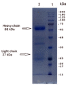
Figure 1
Figure 1
IgY purified from chicken egg yolk was resolved on 12% SDS-PAGE gels and visualized by staining with Coomassie Brilliant Blue. Two major protein bands with molecular weights of 68 kDa and 27 kDa, representing the heavy and light chains of IgY, respectively, were detected (arrows).
Figure 2
Figure 2
Kinetics of serum and egg yolk anti-MERS COV-S IgY antibodies response of chickens after immunization with MERS COV-S recombinant protein compared with the adjuvant-immunized chicken (adjuvant control). Each week is represented by a pool of egg yolks of individual chicken in each group (S1-immunized and adjuvant-immunized).
Figure 3
Figure 3
Western blot analysis of anti-MERS-COV rS1 IgY antibodies. (Left) The S1 protein of MERS-COV was subjected to SDS-PAGE under reducing conditions; (Right) Western blot analysis of the anti-S1 IgY antibody response. SDS gels were electrically transferred onto nitrocellulose membranes and probed with IgY from immunized and nonimmunized hens (marker: molecular maker; lane A: S1-immunized IgY; lane B: adjuvant-immunized IgY). The strips were processed separately and pasted beside each other for documentation.
Figure 4
Figure 4
Dot blotting analysis. Purified anti-S1 IgY antibodies showed reactivity with different concentrations of the spike protein (S), S1, and receptor binding domain (RBD), but had no reactivity with nucleocapsid (NP) protein of MERS CoV.
Figure 5
Figure 5
Recognition by anti-S1 IgY antibodies of viral antigen expressed in MERS-CoV-infected Vero E6 cells, using indirect immunofluorescence assay. (A) Vero E6 cells inoculated with MERS-CoV and stained with anti-S1 IgY antibodies and FITC-conjugated anti-chicken antibodies; and (B) control adjuvant IgY (Bright-field).
Figure 6
Figure 6
Examples of different concentrations of anti-S1 IgY antibodies tested against MERS-CoV on Vero-E6 cells examined by CPE. The IC100 neutralization of the antibody were determined as the reciprocal of the highest dilution at which no CPE was observed.
Figure 7
Figure 7
Evaluation of the neutralizing potential of anti-S1 IgY antibodies, using plaque reduction neutralization test. (A) MERS-CoV (MOI 0.01) was incubated with different concentrations of anti-S1 IgY antibodies and added to Vero E6 cells. After virus adsorption, agar medium was added to the Vero E6 cells, and the plaques that formed were stained with crystal violet, each IgY concentration was tested in triplicate. (B) Percent inhibition of anti-S1 IgY antibodies with different concentrations. The best fit equation is:
Figure 8
Figure 8
(A) Viral titer in the lungs of MERS-CoV mice treated with anti-SI IgY antibodies and control IgY (adjuvant). (B) Body weight changes after MERS-CoV infection between anti-SI IgY antibodies and IgY of adjuvant control group. (CF) Histopathology of the lungs from human dipeptidyl peptidase 4 (hDPP4)-transgenic mice on day 8 after inoculation with MERS-CoV. Representative histopathological findings of mice with the highest cellular infiltration in alveoli by H&E staining (C) Massive mononuclear cell infiltrations including macrophages and lymphocytes with regenerated type II pneumocytes were seen in adjuvant control group (right column), but less in the anti-S1 IgY treated group (left column). Scale bars: 200 μm (upper row) and 20 μm (lower row). Al, alveoli; Br, bronchi; V, vessel. Detection of viral antigen in lung tissues of mice by immunohistochemistry (D) A few antigen positive cells were seen in the lungs of anti-S1 IgY treated group compared to adjuvant control group. Quantification of inflammation areas (E) The area of pulmonary lesion was determined based on the mean percentage of affected areas in each section of the collected lobes form each animal (n = 8 or 6). Circles indicate averages from three observation lobes in each mouse. p = 0.1709 by Mann-Whitney test. Numbers of viral antigen positive cells in the alveoli (F) Data were obtained from 8 or 6 mice. Circles indicate averages of 5 observation fields in each mouse. * p = 0.0196 by Mann-Whitney test.

References

    1. Khomich O.A., Kochetkov S.N., Bartosch B., Ivanov A.V. Redox Biology of Respiratory Viral Infections. Viruses. 2018;10:392. doi: 10.3390/v10080392. - DOI - PMC - PubMed
    1. Widjaja I., Wang C., van Haperen R., Gutierrez-Alvarez J., van Dieren B., Okba N.M.A., Raj V.S., Li W., Fernandez-Delgado R., Grosveld F., et al. Towards a solution to MERS: Protective human monoclonal antibodies targeting different domains and functions of the MERS-coronavirus spike glycoprotein. Emerg. Microbes Infect. 2019;8:516–530. doi: 10.1080/22221751.2019.1597644. - DOI - PMC - PubMed
    1. Zaki A.M., van Boheemen S., Bestebroer T.M., Osterhaus A.D., Fouchier R.A. Isolation of a novel coronavirus from a man with pneumonia in Saudi Arabia. N. Engl. J. Med. 2012;367:1814–1820. doi: 10.1056/NEJMoa1211721. - DOI - PubMed
    1. Zumla A., Hui D.S., Perlman S. Middle East respiratory syndrome. Lancet. 2015;386:995–1007. doi: 10.1016/S0140-6736(15)60454-8. - DOI - PMC - PubMed
    1. World Health Organization Mers Situation Update. [(accessed on 30 October 2020)]; Available online: http://www.emro.who.int/health-topics/mers-cov/mers-outbreaks.html.

LinkOut - more resources