Review
. 2020 Nov 2;5(1):256.
doi: 10.1038/s41392-020-00373-7.

COVID-19-associated gastrointestinal and liver injury: clinical features and potential mechanisms

Affiliations
Review

COVID-19-associated gastrointestinal and liver injury: clinical features and potential mechanisms

Peijie Zhong et al. Signal Transduct Target Ther. .

Abstract

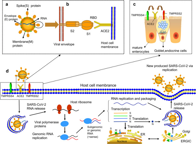
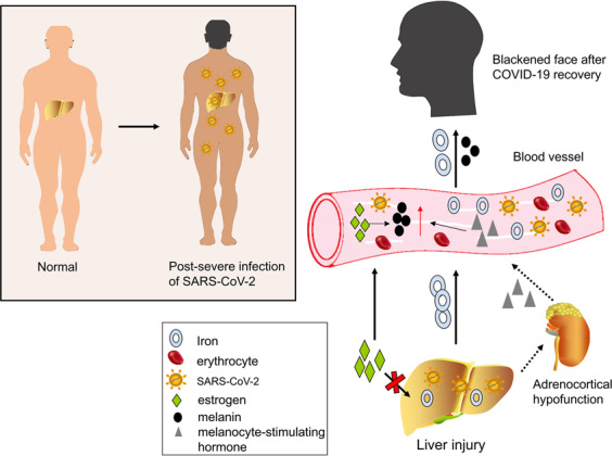
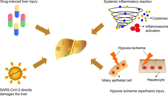
Coronavirus disease-2019 (COVID-19) is caused by severe acute respiratory syndrome coronavirus 2 (SARS-CoV-2). The infection is spreading globally and poses a huge threat to human health. Besides common respiratory symptoms, some patients with COVID-19 experience gastrointestinal symptoms, such as diarrhea, nausea, vomiting, and loss of appetite. SARS-CoV-2 might infect the gastrointestinal tract through its viral receptor angiotensin-converting enzyme 2 (ACE2) and there is increasing evidence of a possible fecal-oral transmission route. In addition, there exist multiple abnormalities in liver enzymes. COVID-19-related liver injury may be due to drug-induced liver injury, systemic inflammatory reaction, and hypoxia-ischemia reperfusion injury. The direct toxic attack of SARS-CoV-2 on the liver is still questionable. This review highlights the manifestations and potential mechanisms of gastrointestinal and hepatic injuries in COVID-19 to raise awareness of digestive system injury in COVID-19.


Conflict of interest statement

The authors declare no competing interests.

Figures

Fig. 1
Fig. 1
Proposed model of SARS-CoV-2 structure and the life cycle of SARS-CoV-2 in host cells. a The structure of SARS-CoV-2. b The entry of SARS-CoV-2 into host cells. Transmembrane spike (S) glycoprotein forms homotrimers protruded from the surface of SARS-CoV-2 to recognize human host ACE2 protein. Receptor-binding domain (RBD) is directly involved in the recognition process. c TMPRSS2 and TMPRSS4, two mucose-specific serine proteases, can promote the infection of SARS-CoV-2 on ACE2+ intestinal epithelial cells. TMPRSS4 is higher expressed than TMPRSS2 in mature enterocytes, while TMPRSS2 is higher expressed than TMPRSS4 in goblet, endocrine cells. d Life cycle of SARS-CoV-2 in host cells. First, S protein of SARS-CoV-2 is combined with ACE2 to form ACE2–virus complex. SARS-CoV-2 is transported to host cells with the assistance of TMPRSS2 and TMPRSS4. Second, SARS-CoV-2 RNA is released into host cytoplasm. SARS-CoV-2 RNA conducts translation of viral polymerase proteins via host ribosome. Third, the negative (−)-sense genomic RNA is synthesized and guide synthesis of subgenomic or genomic positive (+)-sense RNA. Nucleocapsids of SARS-CoV-2 are assembled from genomic RNA and N proteins. Other structures of SARS-CoV-2 such as spike (S) protein, envelope (E) protein and membrane (M) protein are translated in the host endoplasmic reticulum (ER). Finally, the viral RNA-N complex and S, M, and E proteins enter ERGIC (endoplasmic reticulum (ER)–Golgi intermediate compartment) and produce a completely new SARS-CoV-2. The new produced SARS-CoV-2 is released from the host cell through exocytosis
Fig. 2
Fig. 2
Patients with severe COVID-19 develop facial blackness and dull skin after recovery. Liver injury during COVID-19 is mainly responsible for these special manifestations. Three possible mechanisms are presented: (1) iron in the damaged liver drains into blood vessels. The blood with high iron level can lead to a blackening of the face once it supplies to the facial skin; (2) estrogen cannot be metabolized in the damaged liver. An increase in estrogen in the blood eventually causes an increase in conversion of tyrosine to melanin; (3) when liver function is impaired, adrenocortical function is hypoactive and melanocyte-stimulating hormone increases
Fig. 3
Fig. 3
Mechanisms of COVID-19-associated liver injury: (1) drug-induced liver injury; (2) systemic inflammatory response (inflammatory cytokine storm); (3) hypoxic ischemia–reperfusion injury; (4) direct toxic effect of SARS-CoV-2 on the liver

References

    1. World Health Organization. Coronavirus disease (COVID-19) pandemic, 2020. https://www.who.int/health-topics/coronavirus#tab=tab_1 (2020).
    1. Parasa S, et al. Prevalence of gastrointestinal symptoms and fecal viral shedding in patients with coronavirus disease 2019: a systematic review and meta-analysis. JAMA Netw. Open. 2020;3:e2011335. doi: 10.1001/jamanetworkopen.2020.11335. - DOI - PMC - PubMed
    1. Holshue ML, et al. First case of 2019 novel coronavirus in the United States. N. Engl. J. Med. 2020;382:929–936. doi: 10.1056/NEJMoa2001191. - DOI - PMC - PubMed
    1. Guan WJ, et al. Clinical characteristics of coronavirus disease 2019 in China. N. Engl. J. Med. 2020;382:1708–1720. doi: 10.1056/NEJMoa2002032. - DOI - PMC - PubMed
    1. Wang Q, et al. Pattern of liver injury in adult patients with COVID-19: a retrospective analysis of 105 patients. Mil. Med. Res. 2020;7:28. - PMC - PubMed

Publication types

MeSH terms

Substances