. 2020 Nov 2;11(1):5541.
doi: 10.1038/s41467-020-19347-y.

Construction of a complete set of Neisseria meningitidis mutants and its use for the phenotypic profiling of this human pathogen

Affiliations

Construction of a complete set of Neisseria meningitidis mutants and its use for the phenotypic profiling of this human pathogen

Alastair Muir et al. Nat Commun. .

Abstract

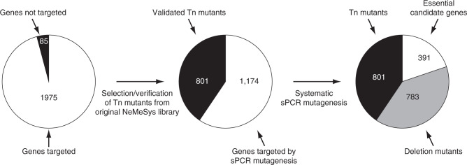
The bacterium Neisseria meningitidis causes life-threatening meningitis and sepsis. Here, we construct a complete collection of defined mutants in protein-coding genes of this organism, identifying all genes that are essential under laboratory conditions. The collection, named NeMeSys 2.0, consists of individual mutants in 1584 non-essential genes. We identify 391 essential genes, which are associated with basic functions such as expression and preservation of genome information, cell membrane structure and function, and metabolism. We use this collection to shed light on the functions of diverse genes, including a gene encoding a member of a previously unrecognised class of histidinol-phosphatases; a set of 20 genes required for type IV pili function; and several conditionally essential genes encoding antitoxins and/or immunity proteins. We expect that NeMeSys 2.0 will facilitate the phenotypic profiling of a major human bacterial pathogen.


Conflict of interest statement

The authors declare no competing interests.

Figures

Fig. 1
Fig. 1. Flowchart of the construction of the NeMeSys 2.0 complete collection of mutants in N. meningitidis 8013.
As for similar efforts in other bacteria,,, we first selected protein-coding genes to be targeted by systematic mutagenesis, excluding 85 genes (4.1%, highlighted in black in the first pie chart) because they encode transposases of repeated insertion sequences, or correspond to short remnants of truncated genes or cassettes (Supplementary Data 1). We then followed a two-step mutagenesis approach explained in the text and in Supplementary Fig. 1. In brief, we first selected a subset of sequence-verified Tn mutants from a previously constructed arrayed library,. Mutations were re-transformed in strain 8013 and PCR-verified. We thus selected 801 Tn mutants with a disrupting transposon in the corresponding target genes (highlighted in black in the second and third pie charts). Next, we systematically mutagenised the remaining 1174 target genes using a validated no-cloning mutagenesis method relying on sPCR. For each successful transformation, two colonies were isolated and PCR-verified. To minimise false-positive identification of essential genes, each transformation that yielded no transformants was repeated at least three times. In total, we could construct an additional 783 mutants (highlighted in grey in the third pie chart), generating an ordered library of defined mutants in 1584 meningococcal genes (Supplementary Data 3). This effort also identified 391 candidate essential genes, which could not be disrupted, encoding proteins required for N. meningitidis growth on rich medium (Supplementary Data 4).
Fig. 2
Fig. 2. Partition of essential meningococcal genes into persistent, shell and cloud genomes.
To perform this analysis, we used the PPanGGOLiN method as explained in the text. a Partition of the 2060 genes in the genome of N. meningitidis 8013: persistent (not highlighted), shell (highlighted in grey), and cloud (highlighted in black). The corresponding datasets are listed in Supplementary Data 5. b Partition of the subset of 391 essential genes identified in this study (same colour code than in a). The corresponding datasets are listed in Supplementary Data 5.
Fig. 3
Fig. 3. Comparison of N. meningitidis essential genome to that of other bacteria.
a Edwards–Venn diagram displaying overlaps between essential genes in bacteria in which complete libraries of mutants have been constructed: N. meningitidis (black line), E. coli (green line), A. baylyi (red line) and S. sanguinis (blue line). To perform this analysis, we queried the DEG database of essential genes using our set of essential genes. The corresponding datasets are listed in Supplementary Data 6. b Comparison of N. meningitidis essential genome (black line) to JCVI-syn3.0 (red line), a synthetic M. mycoides designed with a minimal genome. The corresponding datasets are listed in Supplementary Data 7.
Fig. 4
Fig. 4. Concise cellular overview of the essential meningococcal genome.
Essential genes were integrated into networks and metabolic pathways using primarily MetaCyc. The four basic functional groups are highlighted using the same colour code as in Supplementary Fig. 2, i.e. orange (gene/protein expression), green (genome/cell replication), blue (cell membrane/wall biogenesis) and yellow (cytosolic metabolism). The 34 essential genes that could not be clearly assigned to one of these four categories are not represented on the figure. Genes involved in the different reactions are indicated by their name or NMV_ label, in red when essential, in black when dispensable. *Genes involved in more than one pathway. Key compounds/proteins are abbreviated as follows. ACP acyl carrier protein, AIR aminoimidazole ribotide, CDP-DAG CDP-diacylglycerol, CMP-Kdo CMP-ketodeoxyoctonate, DHAP dihydroxyacetone phosphate, DPP dimethylallyl diphosphate, DXP 1-deoxyxylulose-5P, Fe-S iron-sulfur, FMN flavin mononucleotide, G3P glyceraldehyde-3P, Glu-6P glucose-6P, IMP inosine monophosphate, IPP isopentenyl diphosphate, LOS lipo-oligosaccharide, M-DAP meso-diaminopimelate, NaMN nicotinate d-ribonucleotide, OPP all-trans-octaprenyl diphosphate, PE phosphatidylethanolamine, PG phosphatidylglycerol, PHBA p-hydroxybenzoate, PRPP 5-phosphoribosyl diphosphate, Rib-5P ribulose-5P, SAM S-adenosyl-methionine, UDP-GlcNAc UDP-N-acetyl-glucosamine, UPP di-trans-poly-cis-undecaprenyl diphosphate. The corresponding datasets are listed in Supplementary Data 8.
Fig. 5
Fig. 5. Essential genes in RGP—NMV_1479 and NMV_0559—are conditionally essential.
a Gene organisation of the putative antitoxin NMV_1478 (highlighted in red) with its neighbouring NMV_1479 toxin. Results of the mutagenesis (represented by scissors) are shown. Viable mutant (represented by a grey diplococcus); lethal phenotype (represented by a skull). b Gene organisation of the tps RGP (RGP_0) to which NMV_0559 (highlighted in red) belongs (Supplementary Data 9). Genes on the + strand are in white, genes on the—strand are in black. Results of the mutagenesis (scissors) are shown. Viable mutant (grey diplococcus); lethal phenotype (skull). In contrast to NMV_0559, each of the other target genes in the tps RGP could be mutated individually (not shown for readability).
Fig. 6
Fig. 6. NMV_1317 encodes a novel histidinol-phosphatase.
a Histidine biosynthesis pathway in the meningococcus, with genes highlighted in red. PRPP 5-phosphoribosyl diphosphate, PRFAR phosphoribulosylformimino-AICAR-P, IGP erythro-imidazole-glycerol-P. b Growth on M9 minimal medium, with or without added histidine (His). The plates also contained 0.5 mM IPTG for inducing expression of the complementing genes. WT, strain 8013; Δ1317, ΔNMV_1317 mutant; Δ1317::1317, ΔNMV_1317 complemented with NMV_1317; Δ1317::hisBEC, ΔNMV_1317 cross-complemented with hisBEC from E. coli, which encodes the unrelated histidinol-phosphatase present in this species; Δ1718, ΔNMV_1718 mutant; Δ1718::hisBEC, control showing that ΔNMV_1718 (hisH) cannot be cross-complemented by hisBEC. Source data are provided as a Source Data file.
Fig. 7
Fig. 7. Assaying piliation in the mutants in genes not previously associated with T4P biology in N. meningitidis.
The WT strain and a non-piliated ΔpilD mutant were included as positive and negative controls, respectively. ΔtsaP, ΔtsaP mutant; Δ1205, ΔNMV_1205 mutant; Δ2228, ΔNMV_2228 mutant. a T4P purified using a shearing/precipitation method were separated by SDS-PAGE and either stained with Coomassie blue (upper panel) or analysed by immunoblotting using an antibody against the major pilin PilE (lower panel). Samples were prepared from equivalent numbers of cells and identical volumes were loaded in each lane. MW molecular weight marker lane, with values in kDa. Source data are provided as a Source Data file. b T4P were quantified by whole-cell ELISA using a monoclonal antibody specific for the filaments of strain 8013. Equivalent numbers of cells, based on OD600 readings, were applied to the wells of microtiter plates. Results are expressed in % piliation (ratio to WT) and are the average ± standard deviations from five independent experiments. Source data are provided as a Source Data file. For statistical analysis, one-way ANOVA followed by Dunnett’s multiple comparison tests were performed (****P < 0.0001).
Fig. 8
Fig. 8. Functional analysis of the mutants in genes not previously associated with T4P biology in N. meningitidis.
The WT strain and a non-piliated pilD mutant were included as positive and negative controls, respectively. a Aggregation in liquid culture as assessed by phase-contrast microscopy. ΔtsaP::tsaP, ΔtsaP complemented with tsaP; ΔtsaPpilT, double mutant in tsaP and pilT; Δ1025::1205, ΔNMV_1205 complemented with NMV_1205; Δ1205/ΔpilT, double mutant in NMV_1205 and pilT; Δ2228::2228, ΔNMV_2228 complemented with NMV_2228. Scale bar, 20 µm. Source data are provided as a Source Data file. b Quantification of the competence for DNA transformation. Equivalent numbers of recipient cells were transformed using a rpoB PCR product containing a point mutation leading to rifampicin resistance. Results are expressed as transformation frequencies and are the average ± standard deviations from four independent experiments. Source data are provided as a Source Data file. For statistical analysis, one-way ANOVA followed by Dunnett’s multiple comparison tests were performed (****P < 0.0001).

References

    1. Jolley KA, Maiden MC. BIGSdb: Scalable analysis of bacterial genome variation at the population level. BMC Bioinformatics. 2010;11:595. doi: 10.1186/1471-2105-11-595. - DOI - PMC - PubMed
    1. Ghatak S, King ZA, Sastry A, Palsson BO. The y-ome defines the 35% of Escherichia coli genes that lack experimental evidence of function. Nucleic Acids Res. 2019;47:2446–2454. doi: 10.1093/nar/gkz030. - DOI - PMC - PubMed
    1. Galperin MY, Koonin EV. From complete genome sequence to ‘complete’ understanding? Trends Biotechnol. 2010;28:398–406. doi: 10.1016/j.tibtech.2010.05.006. - DOI - PMC - PubMed
    1. Akerley BJ, et al. A genome-scale analysis for identification of genes required for growth or survival of Haemophilus influenzae. Proc. Natl Acad. Sci. USA. 2002;99:966–971. doi: 10.1073/pnas.012602299. - DOI - PMC - PubMed
    1. Liberati NT, et al. An ordered, nonredundant library of Pseudomonas aeruginosa strain PA14 transposon insertion mutants. Proc. Natl Acad. Sci. USA. 2006;103:2833–2838. doi: 10.1073/pnas.0511100103. - DOI - PMC - PubMed

Publication types

LinkOut - more resources