. 2020 Nov 2;11(1):5531.
doi: 10.1038/s41467-020-19371-y.

Rapid and accurate determination of atomistic RNA dynamic ensemble models using NMR and structure prediction

Affiliations

Rapid and accurate determination of atomistic RNA dynamic ensemble models using NMR and structure prediction

Honglue Shi et al. Nat Commun. .

Abstract

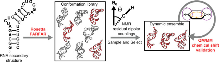
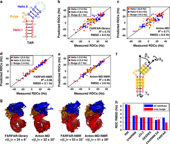
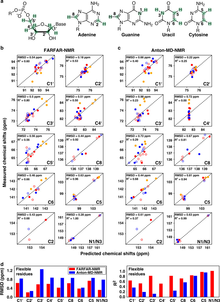
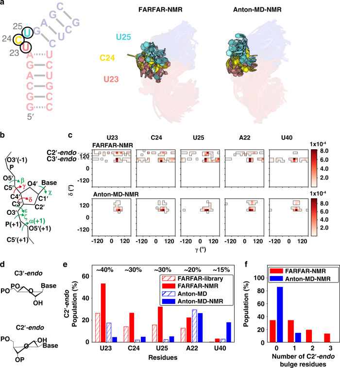
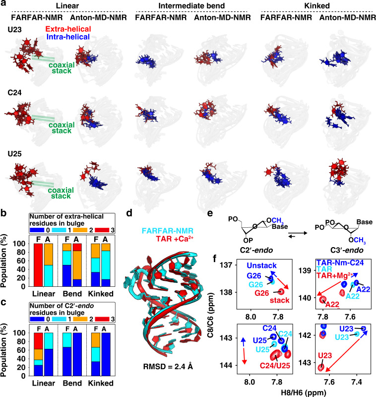
Biomolecules form dynamic ensembles of many inter-converting conformations which are key for understanding how they fold and function. However, determining ensembles is challenging because the information required to specify atomic structures for thousands of conformations far exceeds that of experimental measurements. We addressed this data gap and dramatically simplified and accelerated RNA ensemble determination by using structure prediction tools that leverage the growing database of RNA structures to generate a conformation library. Refinement of this library with NMR residual dipolar couplings provided an atomistic ensemble model for HIV-1 TAR, and the model accuracy was independently supported by comparisons to quantum-mechanical calculations of NMR chemical shifts, comparison to a crystal structure of a substate, and through designed ensemble redistribution via atomic mutagenesis. Applications to TAR bulge variants and more complex tertiary RNAs support the generality of this approach and the potential to make the determination of atomic-resolution RNA ensembles routine.


Conflict of interest statement

H.M.A. is an advisor to and holds an ownership interest in Nymirum, an RNA-based drug discovery company. The remaining authors declare no competing interests.

Figures

Fig. 1
Fig. 1. The FARFAR-NMR pipeline.
An RNA conformation library is generated by Rosetta FARFAR structure prediction using an RNA secondary structure. The FARFAR-NMR ensemble can be refined from the conformation library using NMR RDCs and cross-validated by computing NMR chemical shifts using QM/MM calculations.
Fig. 2
Fig. 2. Using FARFAR-NMR to determine a TAR ensemble.
a Secondary structure of TAR. be Comparison between measured and predicted TAR RDCs for the b FARFAR-library (N = 10,000), c Anton-MD library (N = 10,000), d FARFAR-NMR (N = 20), and e Anton-MD-NMR (N = 20) ensembles. Values in parentheses denote the RDC RMSD. RDCs are color-coded according to the structural elements in a. f Definition of the three inter-helical Euler angles (αh, βh, γh) describing the relative orientation of the two helices (“Methods”). The bps of the upper and lower helices used for defining the Euler angles are highlighted in yellow. g Structural overlay of the TAR libraries and NMR refined ensembles (N = 20) (“Methods”). |βh| is the average absolute magnitude of the bend angle. h Comparison of RDC RMSD overall (blue) or bulge (red) residues for ensembles generated using different MD force fields (ff99, CHARMM36, ff99bsc0χOL3, and modified ff14 DESRES), multiple simulations using a simulated annealing approach (CHARMM SA), and FARFAR.
Fig. 3
Fig. 3. Evaluating TAR ensembles using chemical shifts.
a Chemical structures of the sugar and base moieties with chemical shift probes used to test ensemble accuracy highlighted in green. b, c Comparison of measured and predicted 13C/15N chemical shifts for b FARFAR-NMR (N = 20) and c Anton-MD-NMR (N = 20) by AF-QM/MM (“Methods”). Values are color-coded according to the structural elements in Fig. 2a. Chemical shifts for central Watson–Crick bps within A-form helices (C19-G43, A20-U42, G21-C41, A27-U38, G28-C37) are denoted using open circles. A correction was applied to the predicted chemical shifts (“Methods”) as described previously. For 1H chemical shifts see Supplementary Fig. 3. d Comparison of RMSD (left) and R2 (right) between measured and predicted 13C/15N chemical shifts for flexible residues (U23, C24, U25, A22-U40, G26-C39, C29-G36, G18-C44) for FARFAR-NMR (red) and Anton-MD-NMR (blue).
Fig. 4
Fig. 4. FARFAR-NMR ensemble broadly samples non-canonical sugar-backbone torsion angles relative to Anton-MD-NMR.
a Overlay of the dynamic ensembles of the TAR bulge. b RNA backbone torsion angles exhibiting different and similar distributions between Anton-MD-NMR and FARFAR-NMR are colored red and green, respectively (“Methods”). c 2D density maps of δ versus γ comparing Anton-MD-NMR and FARFAR-NMR ensembles (N = 2000) for bulge residues as well as A22 and U40. The bin width is 20°. d Structure of the ribose moiety in C3′-endo and C2′-endo conformations. e Population of C2′-endo pucker at bulge residues as well as A22 and U40 in the FARFAR-library (N = 10,000, red open), FARFAR-NMR (N = 20 × 100 = 2000, red fill), Anton-MD library (N = 10,000, blue open) and Anton-MD-NMR (N = 20 × 100 = 2000, blue fill). Experimental estimates of the C2′-endo population based on 13C chemical shifts are indicated above the bars (“Methods”). f The population of conformers in the ensemble as a function of the number of C2′-endo bulge residues for FARFAR-NMR (red, N = 20 × 100 = 2000) and Anton-MD-NMR (blue, N = 20 × 100 = 2000).
Fig. 5
Fig. 5. Cooperative extra-helical flipping and sugar repuckering of bulge residues are coupled to coaxial stacking.
a Overlay of conformers showing motions in bulge residues for linear (|βh| < 45°), intermediate bend (45° < |βh| < 70°) and kinked (|βh| > 70°) inter-helical conformations in the FARFAR-NMR and Anton-MD-NMR ensembles. b, c The fractional populations of conformers with the number of b extra-helical and c C2′-endo residues (color-coded) in the bulge residues (U23, C24, and U25) as a function of bending angle in the FARFAR-NMR (“F”) and Anton-MD-NMR (“A”) ensembles (N = 20). d Comparison of one coaxially stacked FARFAR-NMR conformer with the crystal structure of Ca2+-bound TAR (PDBID: 397D). e Nm shifts sugar-pucker equilibrium towards C3′-endo. f Overlay of 2D [13C, 1H] HSQC NMR spectra of the aromatic spins for TAR-Nm-C24 without Mg2+ (blue) with blue arrows indicate unstacking, TAR without Mg2+ (cyan) and TAR with 3 mM Mg2+ (red) with red arrows indicating increased coaxial stacking.
Fig. 6
Fig. 6. Dynamic ensembles of TAR and its bulge variants in the absence and presence of 3 mM Mg2+.
a Secondary structure of TAR and its bulge variants (U1-TAR, U2-TAR, and U7-TAR). The bps at different helices used for defining the Euler angles are highlighted in yellow. b RDC RMSD for ensembles of TAR variants with ensemble size N obtained using FARFAR-NMR. c Comparison between the average bend angle (<|βh|>) and its standard deviation for the FARFAR-NMR (N = 2000, “Methods”) derived ensembles with the best-fit |βh| values obtained from an order tensor analysis of the RDCs. The error bar in the order tensor analysis corresponds to half the cone radius angle assuming an isotropic model. d, e Distributions of the inter-helical bend angle magnitude |βh| for the ensembles (N = 2000, “Methods”) in the absence d and presence e of Mg2+. f The FARFAR-NMR ensembles of TAR and its bulge variants in the absence (upper) and presence (lower) of 3 mM Mg2+. The ensemble size (N) is labeled below for each TAR bulge variant. Motifs in the ensembles are color-coded as in a.
Fig. 7
Fig. 7. Initial ensembles of various RNA.
a The RNA secondary structure for each RNA from top to bottom: human telomerase P2ab, fluoride riboswitch, preQ1 Class I riboswitch and preQ1 Class II riboswitch. The bps at different helices used for defining the Euler angles are highlighted in yellow. b The NMR structure bundles. c The FARFAR-NMR ensembles with ensemble size (N) labeled below. Different structural elements are color-coded according to a. The average bend angles < |βh|> (N = 10 × 200 = 2000) of the two helices are labeled along with the ensembles. d Comparison of the FARFAR-NMR conformer (cyan) with the closest heavy-atom RMSD to the NMR structure (red).

Similar articles

Cited by

References

    1. Breaker RR. Prospects for riboswitch discovery and analysis. Mol. Cell. 2011;43:867–879. doi: 10.1016/j.molcel.2011.08.024. - DOI - PMC - PubMed
    1. Jarmoskaite I, et al. A quantitative and predictive model for RNA binding by human pumilio proteins. Mol. Cell. 2019;74:966–981. doi: 10.1016/j.molcel.2019.04.012. - DOI - PMC - PubMed
    1. Harcourt EM, Kietrys AM, Kool ET. Chemical and structural effects of base modifications in messenger RNA. Nature. 2017;541:339–346. doi: 10.1038/nature21351. - DOI - PMC - PubMed
    1. Halvorsen M, Martin JS, Broadaway S, Laederach A. Disease-associated mutations that alter the RNA structural ensemble. PLoS Genet. 2010;6:e1001074. doi: 10.1371/journal.pgen.1001074. - DOI - PMC - PubMed
    1. Steinert, H. et al. Pausing guides RNA folding to populate transiently stable RNA structures for riboswitch-based transcription regulation. Elife6, e21297 (2017). - PMC - PubMed

Publication types