

Optical coherence tomography-based deep-learning model for detecting central serous chorioretinopathy
- PMID: 33139813
- PMCID: PMC7608618
- DOI: 10.1038/s41598-020-75816-w
Optical coherence tomography-based deep-learning model for detecting central serous chorioretinopathy
Abstract
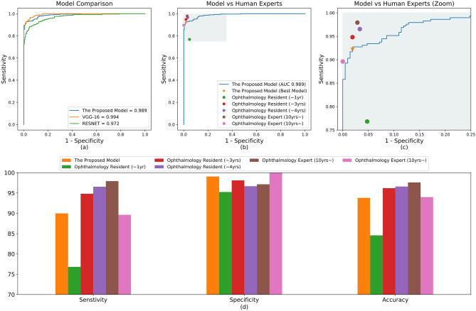
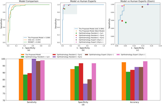
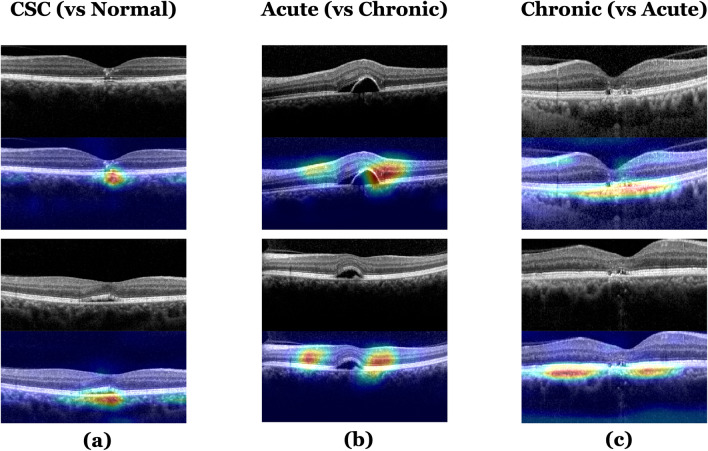
Central serous chorioretinopathy (CSC) is a common condition characterized by serous detachment of the neurosensory retina at the posterior pole. We built a deep learning system model to diagnose CSC, and distinguish chronic from acute CSC using spectral domain optical coherence tomography (SD-OCT) images. Data from SD-OCT images of patients with CSC and a control group were analyzed with a convolutional neural network. Sensitivity, specificity, accuracy, and area under the receiver operating characteristic curve (AUROC) were used to evaluate the model. For CSC diagnosis, our model showed an accuracy, sensitivity, and specificity of 93.8%, 90.0%, and 99.1%, respectively; AUROC was 98.9% (95% CI, 0.983-0.995); and its diagnostic performance was comparable with VGG-16, Resnet-50, and the diagnoses of five different ophthalmologists. For distinguishing chronic from acute cases, the accuracy, sensitivity, and specificity were 97.6%, 100.0%, and 92.6%, respectively; AUROC was 99.4% (95% CI, 0.985-1.000); performance was better than VGG-16 and Resnet-50, and was as good as the ophthalmologists. Our model performed well when diagnosing CSC and yielded highly accurate results when distinguishing between acute and chronic cases. Thus, automated deep learning system algorithms could play a role independent of human experts in the diagnosis of CSC.
Conflict of interest statement
The authors declare no competing interests.
Figures

Similar articles
-
Assessing central serous chorioretinopathy with deep learning and multiple optical coherence tomography images.Sci Rep. 2022 Feb 3;12(1):1831. doi: 10.1038/s41598-022-05051-y. Sci Rep. 2022. PMID: 35115577 Free PMC article.
-
Classifying central serous chorioretinopathy subtypes with a deep neural network using optical coherence tomography images: a cross-sectional study.Sci Rep. 2022 Jan 10;12(1):422. doi: 10.1038/s41598-021-04424-z. Sci Rep. 2022. PMID: 35013502 Free PMC article.
-
Developing and Evaluating an AI-Based Computer-Aided Diagnosis System for Retinal Disease: Diagnostic Study for Central Serous Chorioretinopathy.J Med Internet Res. 2023 Nov 29;25:e48142. doi: 10.2196/48142. J Med Internet Res. 2023. PMID: 38019564 Free PMC article.
-
Morphologic features of large choroidal vessel layer: age-related macular degeneration, polypoidal choroidal vasculopathy, and central serous chorioretinopathy.Graefes Arch Clin Exp Ophthalmol. 2018 Dec;256(12):2309-2317. doi: 10.1007/s00417-018-4143-1. Epub 2018 Sep 27. Graefes Arch Clin Exp Ophthalmol. 2018. PMID: 30259090 Review.
-
[Evaluation of the choroid in central serous chorioretinopathy].Nippon Ganka Gakkai Zasshi. 2012 Nov;116(11):1062-79. Nippon Ganka Gakkai Zasshi. 2012. PMID: 23316655 Review. Japanese.
Cited by
-
Assessing central serous chorioretinopathy with deep learning and multiple optical coherence tomography images.Sci Rep. 2022 Feb 3;12(1):1831. doi: 10.1038/s41598-022-05051-y. Sci Rep. 2022. PMID: 35115577 Free PMC article.
-
Classifying central serous chorioretinopathy subtypes with a deep neural network using optical coherence tomography images: a cross-sectional study.Sci Rep. 2022 Jan 10;12(1):422. doi: 10.1038/s41598-021-04424-z. Sci Rep. 2022. PMID: 35013502 Free PMC article.
-
Differentiating a pachychoroid and healthy choroid using an unsupervised machine learning approach.Sci Rep. 2022 Sep 29;12(1):16323. doi: 10.1038/s41598-022-20749-9. Sci Rep. 2022. PMID: 36175534 Free PMC article.
-
Simple Code Implementation for Deep Learning-Based Segmentation to Evaluate Central Serous Chorioretinopathy in Fundus Photography.Transl Vis Sci Technol. 2022 Feb 1;11(2):22. doi: 10.1167/tvst.11.2.22. Transl Vis Sci Technol. 2022. PMID: 35147661 Free PMC article.
-
Research Progress in Artificial Intelligence for Central Serous Chorioretinopathy: A Systematic Review.Ophthalmol Ther. 2025 Jul 22. doi: 10.1007/s40123-025-01209-9. Online ahead of print. Ophthalmol Ther. 2025. PMID: 40694226 Review.
References
-
- Spaide, R. F. et al. Central serous chorioretinopathy in younger and older adults. Ophthalmology103, 2070–2079; discussion 2079–2080 (1996). - PubMed
-
- Song IS, Shin YU, Lee BR. Time-periodic characteristics in the morphology of idiopathic central serous chorioretinopathy evaluated by volume scan using spectral-domain optical coherence tomography. Am. J. Ophthalmol. 2012;154(366):e364–375.e364. - PubMed
-
- Zhen Y, et al. Assessment of central serous chorioretinopathy depicted on color fundus photographs using deep learning. Retina. 2020;40:1558–1564. - PubMed
Publication types
MeSH terms
LinkOut - more resources
Full Text Sources
