

Absorption cross section of gold nanoparticles based on NIR laser heating and thermodynamic calculations
- PMID: 33139828
- PMCID: PMC7606525
- DOI: 10.1038/s41598-020-75895-9
Absorption cross section of gold nanoparticles based on NIR laser heating and thermodynamic calculations
Abstract
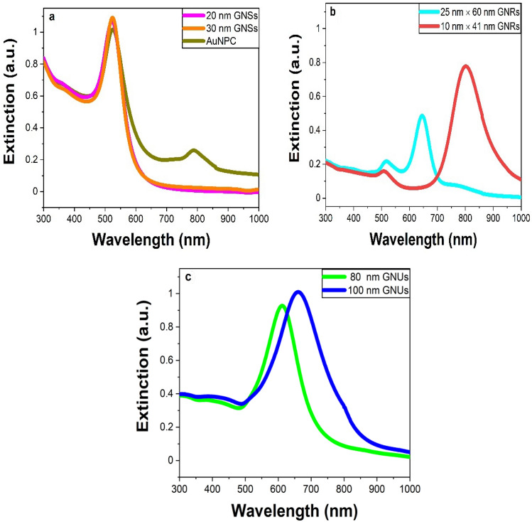
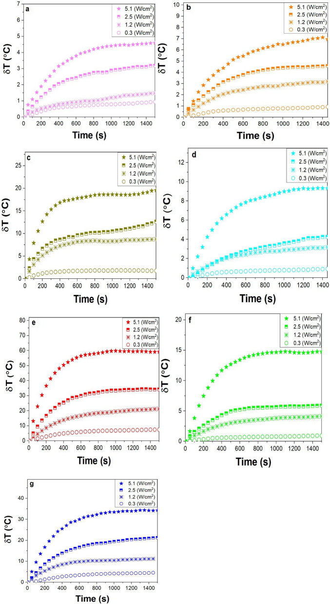
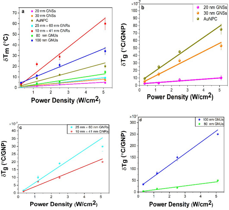
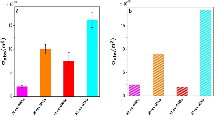
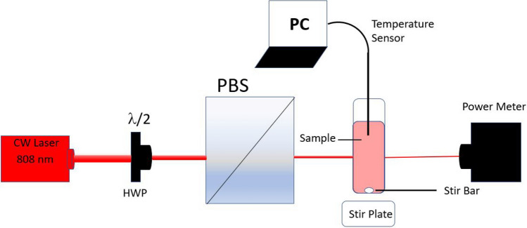
We present a method for measuring the optical absorption cross section ([Formula: see text]) of gold nanoparticles (GNPs) based on optically heating the solution of GNPs with an 808 nm near-infrared (NIR) laser and measuring the temperature increase of the solution. We rely on the theoretical calculations based on the heat diffusion equations and experimental measurements based on the energy balance equations to measure the [Formula: see text] and the temperature distribution of single GNPs. Several morphologies, including gold nanospheres (GNSs), spherical gold nanoparticle conjugate (AuNPC), which are 20 nm GNSs surface-functionalized with an IR 808 dye, gold nanorods (GNRs), and gold nanourchins (GNUs), were studied. The study found that a single 20 nm GNS has the lowest [Formula: see text] and temperature distribution as compared to 100 nm GNUs. By increasing the size of GNSs from 20 to 30 nm, the magnitude of [Formula: see text] as well as temperature distribution increases by a factor of 5. The [Formula: see text] values of 20 and 30 nm GNSs calculated by Mie theory and the experimentally measured are in a good agreement. GNRs with equivalent radius ([Formula: see text]) 9.16 nm show the second lowest [Formula: see text]. By increasing the [Formula: see text] by a factor of 2 to 19.2 nm, the measured [Formula: see text] and temperature distribution also increased by a factor of 2. We also estimated [Formula: see text] for GNUs with diameters at 80 and 100 nm, which also have higher [Formula: see text] values. This work confirms that we can use temperature to accurately measure the [Formula: see text] of a variety of GNPs in solution.
Conflict of interest statement
The authors declare no competing interests.
Figures
References
-
- Cole JR, et al. Photothermal efficiencies of nanoshells and nanorods for clinical therapeutic applications. J. Phys. Chem. C. 2009;113:12090–12095. doi: 10.1021/jp9003592. - DOI
LinkOut - more resources
Full Text Sources
Other Literature Sources
Miscellaneous
