. 2021 Apr;18(4):936-944.
doi: 10.1038/s41423-020-00573-9. Epub 2020 Nov 2.

Low serum neutralizing anti-SARS-CoV-2 S antibody levels in mildly affected COVID-19 convalescent patients revealed by two different detection methods

Affiliations

Low serum neutralizing anti-SARS-CoV-2 S antibody levels in mildly affected COVID-19 convalescent patients revealed by two different detection methods

Berislav Bošnjak et al. Cell Mol Immunol. 2021 Apr.

Abstract

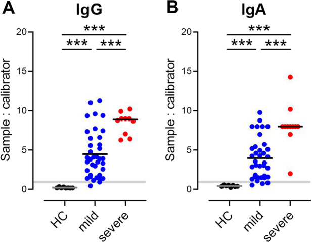
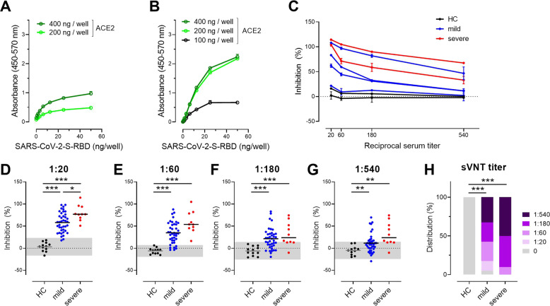
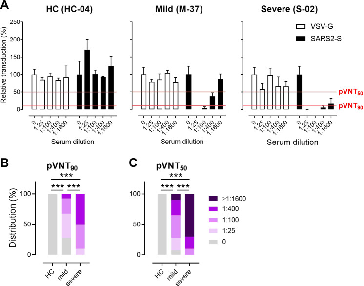
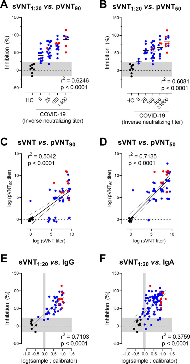
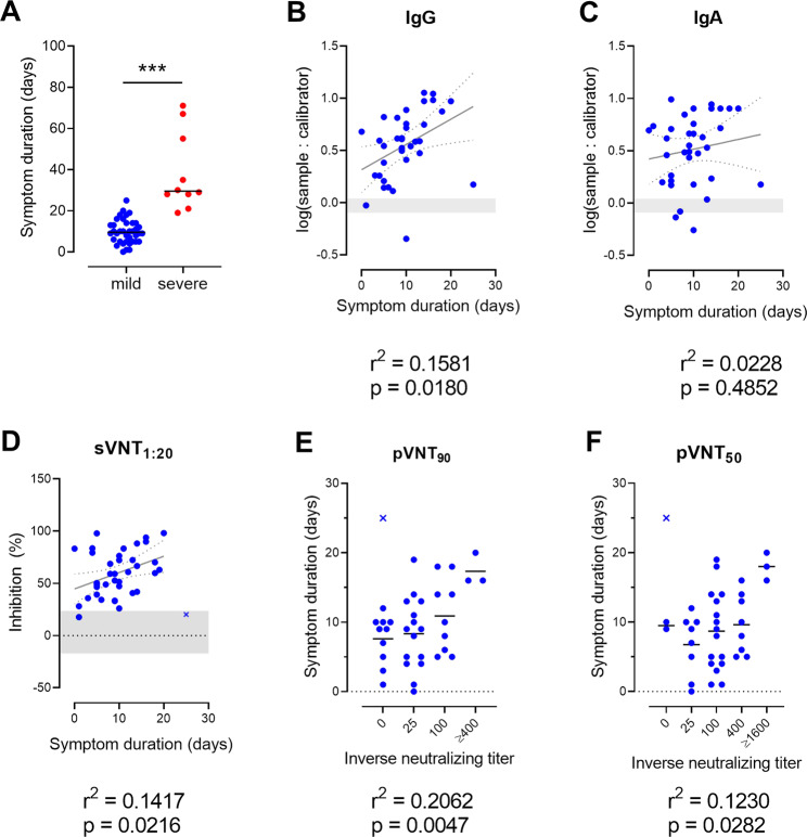
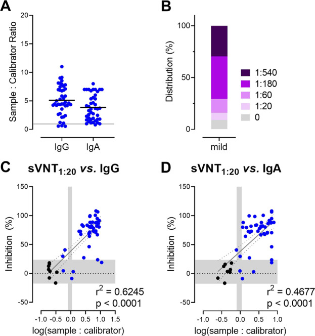
Neutralizing antibodies targeting the receptor-binding domain (RBD) of the SARS-CoV-2 spike (S) block severe acute respiratory syndrome coronavirus 2 (SARS-CoV-2) entry into cells via surface-expressed angiotensin-converting enzyme 2 (ACE2). We used a surrogate virus neutralization test (sVNT) and SARS-CoV-2 S protein-pseudotyped vesicular stomatitis virus (VSV) vector-based neutralization assay (pVNT) to assess the degree to which serum antibodies from coronavirus disease 2019 (COVID-19) convalescent patients interfere with the binding of SARS-CoV-2 S to ACE2. Both tests revealed neutralizing anti-SARS-CoV-2 S antibodies in the sera of ~90% of mildly and 100% of severely affected COVID-19 convalescent patients. Importantly, sVNT and pVNT results correlated strongly with each other and to the levels of anti-SARS-CoV-2 S1 IgG and IgA antibodies. Moreover, levels of neutralizing antibodies correlated with the duration and severity of clinical symptoms but not with patient age. Compared to pVNT, sVNT is less sophisticated and does not require any biosafety labs. Since this assay is also much faster and cheaper, sVNT will not only be important for evaluating the prevalence of neutralizing antibodies in a population but also for identifying promising plasma donors for successful passive antibody therapy.

Keywords: COVID-19; ELISA; Neutralizing antibody; SARS-CoV-2; Serum.


Conflict of interest statement

The authors declare no competing interests.

Figures

Fig. 1
Fig. 1
Qualitative analysis of serum total IgG (A) and IgA (B) antibodies against SARS-CoV-2 S1 in convalescent patients with mild or severe COVID-19 and healthy controls (HC) determined by ELISA. Shaded area cutoff values to determine positive (above), borderline (within), and negative (below gray area) samples. Dots, individuals; bars, mean, ***P < 0.001, Welch’s ANOVA followed by Dunnett’s T3 multiple comparisons test
Fig. 2
Fig. 2
The surrogate virus neutralization test (sVNT) detects neutralizing antibodies interfering with SARS-CoV-2 S RBD binding to human ACE2. Binding of SARS-CoV-2 S RBD to human ACE2 from commercial vendor (A) and produced in-house (B). Plates were coated with ACE2 as indicated. His-tagged SARS-CoV-2 S RBD was titrated as indicated and detected with an anti-His peroxidase-labeled mAb. Representative assays performed in duplicate are presented as the mean ± SD. C Inhibition of the interaction of SARS-CoV-2 S RBD with ACE2 by the addition of sera from convalescent patients with mild (blue lines) or severe (red lines) COVID-19 and healthy controls (HC; black lines). Assay performed in duplicate; mean percentages of neutralization ± SD. DG Inhibition of the interaction of SARS-CoV-2 S RBD with ACE2 at the serum dilutions indicated. Individual values (dots) and means (line). Shaded areas represent the mean ± 2SD of values from sera of healthy controls. *P < 0.05; ***P < 0.001, Welch’s ANOVA followed by Dunnett’s T3 multiple comparisons test. H Relative distributions of SARS-CoV-2 neutralizing serum titers determined as the dilution retaining binding reduction > mean + 2SD of HC. ***P < 0.001; Fisher’s exact test (HC vs. mild or severe) or the Chi-squared test was used to assess the trend (mild vs. severe)
Fig. 3
Fig. 3
Frequency of neutralizing antibodies against SARS-CoV-2 measured by a pseudotyped virus neutralization test (pVNT) based on SARS-CoV-2 S protein-pseudotyped VSV. A Example of pVNT results. Sera from COVID-19 convalescent patients with mild or severe disease—but not from healthy controls (HC)—suppress entry of replication-defective VSV particles carrying the SARS-CoV-2 S protein into host cells (filled bars); neither sera suppressed the entry of control particles carrying the G-protein of VSV entry (open bars). Red lines indicate levels of 50 or 90% suppression of virus entry as indicated. Relative distributions of SARS-CoV-2 neutralizing serum titers that result in (B) 90% (pVNT90) or (C) 50% (pVNT50) reduction of luciferase production, as described in (A). ***P < 0.001; Fisher’s exact test (HC vs. mild or severe) or the Chi-squared test was used to assess the trend (mild vs. severe)
Fig. 4
Fig. 4
sVNT positively correlates with pVNT and anti-SARS-CoV-2 S1 IgG and IgA antibodies. Correlation between sVNT1:20 and antibody titers resulting in 90% (A) or 50% (B) reduction of luciferase production in pVNT90 and pVNT50. The horizontal shaded area indicates the mean ± 2SD range of inhibition of sera from HC. Correlation between log-transformed sVNT titers (determined as in Fig. 2H) and log-transformed pVNT90 (C) and pVNT50 (D). To alleviate overplotting, the titer values were jittered by the addition of random values in the interval [−0.5, 0.5]. Correlation between sVNT1:20 and log-transformed SARS-CoV-2 S1-specific IgG (E) and IgA (F) levels measured by ELISA. The vertical shaded areas indicate the respective cutoff values recommended by the manufacturer to determine positive (right to), borderline (within) and negative (left to) shaded areas. The horizontal shaded area indicates the mean ± 2SD range of inhibition of sera from healthy controls. AF Dots, samples of HC (black), mildly (blue), or severely (red) affected COVID-19 convalescent cases. CF Linear correlation (solid line) and 95% confidence intervals (dotted lines). Correlation, one-way ANOVA followed by a test for the trend (A, B) or Pearson r (CF)
Fig. 5
Fig. 5
The duration of symptoms correlates with total and neutralizing SARS-CoV-2 S-specific antibody levels. A Symptom duration of mild and severely affected COVID-19 patients. Dots, individuals; bars, mean, ***P < 0.001, Welch’s t test. Weak positive correlation between duration of symptoms and levels of log-transformed SARS-CoV-2 S1-specific IgG (B) and IgA (C) antibodies, sVNT1:20 (D), pVNT90 (E), or pVNT50 (F) neutralizing antibody titers. Dots, convalescent individuals with mild COVID-19, outliers are marked with x, horizontal lines, means. B, C Shaded areas indicate vendor-defined cutoff values to determine positive (above), borderline (within), and negative (below gray area) samples. D The shaded area indicates the mean ± 2SD range of inhibition of sera from HC. Correlation, Pearson r (BD) or one-way ANOVA followed by a test for the trend (E, F). An outlier was defined as a value with absolute residual value > 2SD of all residual values (D) or as a value > mean ± 2SD of values with the same titer
Fig. 6
Fig. 6
sVNT correlates positively with total levels of anti-S IgG and IgA antibodies against SARS-CoV-2 S1 in a validation cohort of convalescent patients with mild COVID-19 (n = 44). A Serum anti-SARS-CoV-2 S1 antibodies determined by ELISA. Shaded areas, cutoff values to determine positive (above), borderline (within), and negative (below gray area) samples. Dots, individuals; bars, mean. B Relative distribution of SARS-CoV-2 neutralizing serum titers determined as the dilution retaining binding reduction > mean + 2SD of HC. Correlation between sVNT1:20 and log-transformed SARS-CoV-2 S1-specific IgG (C) and IgA (D) levels measured by ELISA. Vertical shaded areas, vendor-defined cutoff values to determine positive (right to), borderline (within), and negative (left to shaded area) samples. Horizontal shaded area, the mean ± 2SD inhibition of sera from HC. Correlation, Pearson r

References

    1. Zhu J, et al. Clinical characteristics of 3062 COVID-19 patients: a meta-analysis. J. Med. Virol. 2020;92:1902–1914. doi: 10.1002/jmv.25884. - DOI - PMC - PubMed
    1. Zhu N, et al. A novel coronavirus from patients with pneumonia in China, 2019. N. Engl. J. Med. 2020;382:727–733. doi: 10.1056/NEJMoa2001017. - DOI - PMC - PubMed
    1. Oran DP, Topol EJ. Prevalence of asymptomatic SARS-CoV-2 infection: a narrative review. Ann. Intern. Med. 2020;173:362–367. doi: 10.7326/M20-3012. - DOI - PMC - PubMed
    1. Liu PP, Blet A, Smyth D, Li H. The science underlying COVID-19: implications for the cardiovascular system. Circulation. 2020;142:68–78. doi: 10.1161/CIRCULATIONAHA.120.047549. - DOI - PubMed
    1. Ackermann M, et al. Pulmonary vascular endothelialitis, thrombosis, and angiogenesis in Covid-19. N. Engl. J. Med. 2020;383:120–128. doi: 10.1056/NEJMoa2015432. - DOI - PMC - PubMed