Review
. 2021 Apr;18(4):842-859.
doi: 10.1038/s41423-020-00565-9. Epub 2020 Nov 2.

Tumor-infiltrating lymphocytes in the immunotherapy era

Affiliations
Review

Tumor-infiltrating lymphocytes in the immunotherapy era

Sterre T Paijens et al. Cell Mol Immunol. 2021 Apr.

Abstract

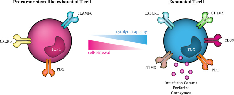
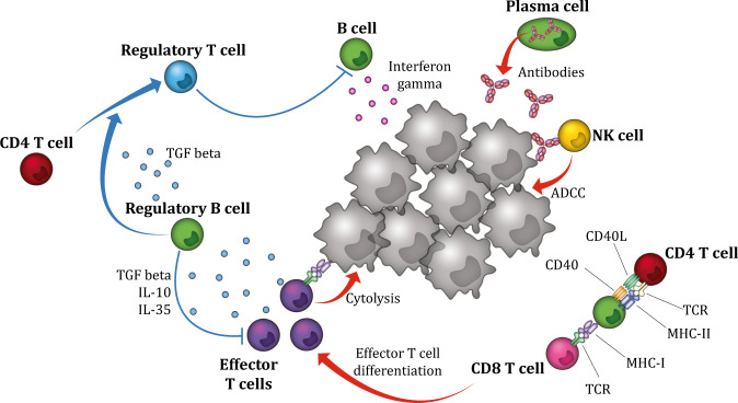
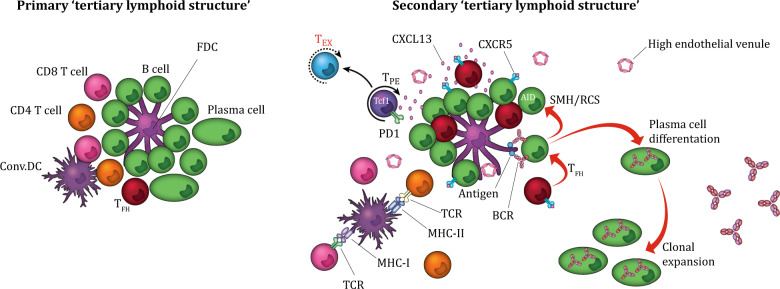
The clinical success of cancer immune checkpoint blockade (ICB) has refocused attention on tumor-infiltrating lymphocytes (TILs) across cancer types. The outcome of immune checkpoint inhibitor therapy in cancer patients has been linked to the quality and magnitude of T cell, NK cell, and more recently, B cell responses within the tumor microenvironment. State-of-the-art single-cell analysis of TIL gene expression profiles and clonality has revealed a remarkable degree of cellular heterogeneity and distinct patterns of immune activation and exhaustion. Many of these states are conserved across tumor types, in line with the broad responses observed clinically. Despite this homology, not all cancer types with similar TIL landscapes respond similarly to immunotherapy, highlighting the complexity of the underlying tumor-immune interactions. This observation is further confounded by the strong prognostic benefit of TILs observed for tumor types that have so far respond poorly to immunotherapy. Thus, while a holistic view of lymphocyte infiltration and dysfunction on a single-cell level is emerging, the search for response and prognostic biomarkers is just beginning. Within this review, we discuss recent advances in the understanding of TIL biology, their prognostic benefit, and their predictive value for therapy.

Keywords: B cells; T cells; Tertiary lymphoid structures; Tumor infiltrating lymphocytes; immunotherapy.


Conflict of interest statement

The authors declare no competing interests.

Figures

Fig. 1
Fig. 1
CD8+ T cell exhaustion states. CD8+ T cell exhaustion is thought of as a gradual process, with various functional and phenotypic states, including TPE and TEX states. The TPE phenotype is characterized by the expression of TCF1, which is lost upon differentiation into TEX cells. Cell surface markers identified on TPE cells include Slamf6, PD1, and CXCR5, and their functional capacity comprises the ability to produce an antigen-specific immune response, persist long-term, self-renew, and eventually differentiate into TEX cells. In contrast, TEX cells expresses mostly coinhibitory cell surface receptors and transcription factors associated with effector and exhausted cells, including CX3CR1, PD1, CD39, and TIM3, which reflects the functional capacity of TEX cells, that is, mostly cytotoxic functions. TEX cells: terminally exhausted T cells, TPE cells: progenitor STEM-like exhausted cells, TCF1: transcription factor 1
Fig. 2
Fig. 2
Antitumor- and protumor-related functional properties of B cells. B cells and plasma cells have several ways to promote local tumor-associated T cell responses. Functionally, B cells may act as antigen-presenting cells and facilitate tumor antigen-derived presentation to T cells. B cells also promote antitumor immunity by secreting immunostimulatory cytokines, such as IFNγ, that drive cytotoxic immune responses. In addition, B cells can directly kill tumor cells by secreting toxic cytokines, such as granzyme B. Plasma cells promote the antitumor immune response by secreting tumor-specific antibodies that can mediate ADCC, resulting in phagocytosis of tumor cells. In contrast, Breg cells suppress the antitumor immune response indirectly by secreting the immunoregulatory cytokines IL-10, IL-35, and TGFβ and directly by inhibiting effector cells, such as cytotoxic CD8+ T cells. Furthermore, Breg cells suppress antitumor immunity by converting CD4+ T cells into Treg cells via TGFβ. ADCC: antibody-dependent cell-mediated cytotoxicity, Breg cells: regulatory B cells, Treg cells: regulatory T cells
Fig. 3
Fig. 3
TLS maturation state and CXCL13. TLSs are optimally organized nodular immune structures consisting of a CD3+ T cell-rich zone with mature DCs in close proximity to CD20+ GC-like follicle B cells intermingled with FDCs and surrounded by plasma cells. The CXCL13–CXCR5 axis regulates the organization of B cells inside the follicles. CXCL13-secreting CD8+ T cells induce chemotaxis by binding to the receptor CXCR5, which is mainly expressed by B cells and TFH cells. Inside the TLS, B cells, T cells, DCs, and FDCs interact and activate each other, promoting a local, sustained, organized immune response. TLS maturation varies from dense lymphocyte aggregates to primary TLSs and secondary follicle-like mature TLSs. The difference between primary and secondary TLSs is the presence of germinal center activity, which is dependent on B cells expressing AID, facilitating SHM and CSR and resulting in high-affinity antibody production by class-switched plasma cells. In addition, mature TLSs are surrounded by HEVs, facilitating lymphocyte migration and extravasation. TLSs: tertiary lymphoid structures, DCs: dendritic cells, FDCs: follicular dendritic cells, TFH cells follicular helper T cells, AID: activation-induced deaminase, SMH: somatic hypermutation, CSR: class switch recombination, HEVs: high endothelial venules

Similar articles

Cited by

References

    1. Zloza A, Al-Harthi L. Multiple populations of T lymphocytes are distinguished by the level of CD4 and CD8 coexpression and require individual consideration. J. Leukoc. Biol. 2006;79:4–6. - PubMed
    1. Parrot T, et al. Transcriptomic features of tumour-infiltrating CD4lowCD8high double positive αβ T cells in melanoma. Sci Rep. 2020;10:5900. - PMC - PubMed
    1. Menard LC, et al. Renal cell carcinoma (RCC) tumors display large expansion of double positive (DP) CD4+CD8+ T cells with expression of exhaustion markers. Front Immunol. 2018;9:2728. - PMC - PubMed
    1. Bohner P, et al. Double positive CD4+CD8+ T cells are enriched in urological cancers and favor T helper-2 polarization. Front Immunol. 2019;10:622. - PMC - PubMed
    1. Desfrançois J, et al. Double positive CD4CD8 αβ T cells: a new tumor-reactive population in human melanomas. PLoS One. 2010;5:e8437. - PMC - PubMed