. 2020 Nov;10(11):2098-2111.
doi: 10.21037/qims-20-607.

Clinical value of dual-phase F-18 sodium fluoride PET/CT for diagnosing bone metastasis in cancer patients with solitary bone lesion

Affiliations

Clinical value of dual-phase F-18 sodium fluoride PET/CT for diagnosing bone metastasis in cancer patients with solitary bone lesion

Jeong Won Lee et al. Quant Imaging Med Surg. 2020 Nov.

Abstract

Background: The present study aimed to investigate whether dual-phase F-18 sodium-fluoride (NaF) positron emission tomography/computed tomography (PET/CT) could improve the diagnostic accuracy of detecting bone metastasis in cancer patients with a solitary bone lesion compared to conventional F-18 NaF PET/CT.

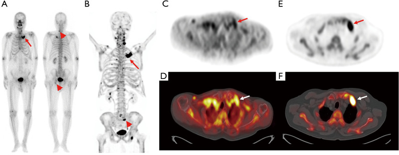
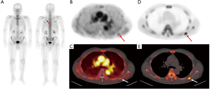
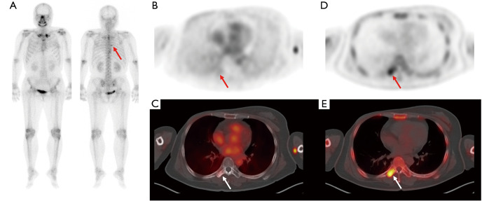
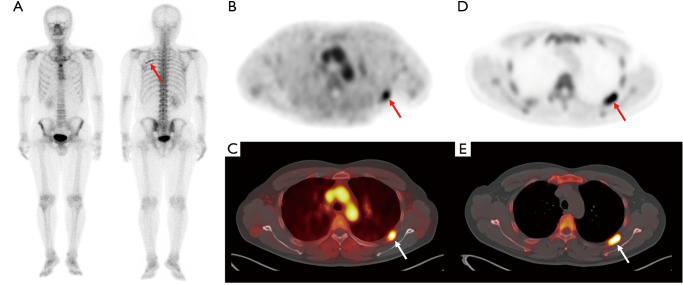
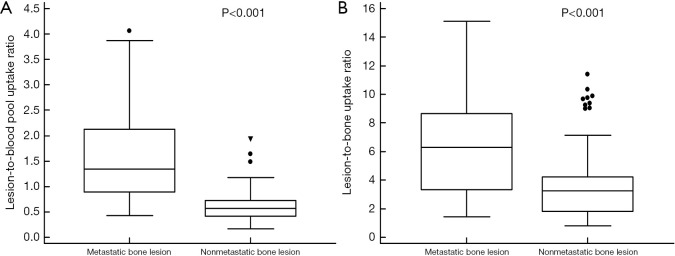
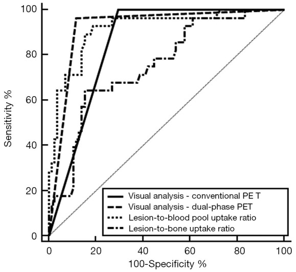
Methods: We retrospectively enrolled 113 cancer patients who underwent dual-phase F-18 NaF PET/CT for the differential diagnosis of a solitary bone lesion seen on bone scintigraphy. According to the dual-phase PET/CT protocol, an early-phase scan was acquired immediately after radiotracer injection and a conventional F-18 NaF PET/CT scan was performed. The diagnostic abilities of the visual analysis of conventional and dual-phase PET/CT scans and two quantitative parameters (lesion-to-blood pool uptake ratio on early-phase scan and lesion-to-bone uptake ratio on conventional scan) for detecting bone metastasis were compared. The final diagnosis of bone metastasis was made by histopathological confirmation or follow-up imaging studies.

Results: A metastatic bone lesion was diagnosed in 28 patients (24.8%). The sensitivity, specificity, and accuracy were 100.0%, 70.6%, and 77.9%, respectively, for visual analysis of conventional F-18 NaF PET/CT, 92.9%, 42.4%, 54.9%, respectively, for lesion-to-bone uptake ratio, 96.4%, 88.2%, and 90.3%, respectively, for visual analysis of dual-phase PET/CT, and 92.9%, 81.2%, and 83.2%, respectively, for lesion-to-blood pool uptake ratio. Visual analysis of dual-phase PET/CT was shown to have the highest area under the receiver operating characteristic curve value (0.923; 95% CI, 0.858-0.965) among all parameters.

Conclusions: Dual-phase F-18 NaF PET/CT showed a high diagnostic ability for detecting bone metastasis with improved specificity and accuracy compared to conventional F-18 NaF PET/CT in cancer patients. Dual-phase F-18 NaF PET/CT might help diagnose bone metastasis in patients with malignancies who were shown to have a solitary bone lesion on bone scintigraphy.

Keywords: Bone; F-18 Sodium-fluoride (NaF); diagnosis; metastasis; positron emission tomography (PET).


Conflict of interest statement

Conflicts of Interest: All authors have completed the ICMJE uniform disclosure form (available at http://dx.doi.org/10.21037/qims-20-607). The authors have no conflicts of interest to declare.

Figures

Figure 1
Figure 1
Bone scintigraphy (A) and transaxial PET (C) and fused PET/CT (D) from the early-phase scan and a maximum intensity projection image (B) and transaxial PET (E) fused PET/CT (F) images from the conventional scan in F-18 NaF PET/CT of a 78-year-old man with prostate cancer. During clinical follow-up after starting hormone treatment, he underwent surveillance bone scintigraphy because of his increased serum prostate-specific antigen level (24.55 ng/mL). On bone scintigraphy images (A), focally increased radiotracer uptake newly appears in his left 1st rib (arrow in A), while focally increased radiotracer uptake in the lower cervical spine and left acetabulum (arrowheads in A) is seen in a previous bone scintigraphy, and considered degenerative bone lesions. Both early-phase and conventional scan images show intensely increased radiotracer uptake at the left 1st rib (arrows in B-F) with a lesion-to-blood pool uptake ratio of 0.94, suggesting metastatic bone lesion. Furthermore, another focally increased radiotracer uptake is observed in the sacrum (arrowhead in B), suggesting another metastatic bone lesion. The left rib lesion was histopathologically confirmed to be a metastatic bone lesion from prostate cancer. PET/CT, positron emission tomography/computed tomography.
Figure 2
Figure 2
Bone scintigraphy (A) and transaxial PET (B) and fused PET/CT (C) from the early-phase scan and transaxial PET (D) and fused PET/CT (E) from the conventional scan in F-18 NaF PET/CT of a 72-year-old man with prostate cancer. Staging bone scintigraphy images show focally increased radiotracer uptake in the left scapula (arrow on A) with serum prostate-specific antigen level of 55.70 ng/mL. Bone-phase scan images show focal intensely increased radiotracer uptake in the left scapula (arrows on D and E); however, only mildly increased radiotracer uptake is observed on early-phase scan images (arrows on B and C) with a lesion-to-blood pool uptake ratio of 0.43. The lesion was clinically diagnosed as being a metastatic bone lesion. PET/CT, positron emission tomography/computed tomography.
Figure 3
Figure 3
Bone scintigraphy (A) and transaxial PET (B) and fused PET/CT (C) from the early-phase scan and transaxial PET (D) and fused PET/CT (E) from the conventional scan in F-18 NaF PET/CT of a 47-year-old woman with breast cancer. On surveillance bone scintigraphy after right mastectomy, focally increased radiotracer uptake is newly observed in the T7 spine (arrow in A). On bone-phase scan images, focal intensely increased radiotracer uptake is shown in the right transverse process of T7 spine (arrows on D and E), but only minimal radiotracer uptake was shown on early-phase scan images (arrows on B and C) with a lesion-to-blood pool uptake ratio of 0.29. On follow-up imaging studies, the bone lesion was clinically diagnosed as nonmetastatic bone lesion. PET/CT, positron emission tomography/computed tomography.
Figure 4
Figure 4
Bone scintigraphy (A) and transaxial (B) and fused PET/CT (C) from the early-phase scan and transaxial PET (D) and fused PET/CT (E) from the conventional scan in F-18 NaF PET/CT of a 39-year-old man with renal cell carcinoma. On surveillance bone scintigraphy performed after curative surgical resection, an elongated rib lesion is shown (arrow on A). The rib lesion shows intensely increased radiotracer uptake on early-phase scan images (arrows on B and C) with a lesion-to-blood pool uptake ratio of 1.48 as well as on bone-phase scan images (arrows on D and E). The rib lesion was clinically diagnosed as nonmetastatic post-traumatic bone lesion by follow-up imaging studies. PET/CT, positron emission tomography/computed tomography.
Figure 5
Figure 5
Distribution of the lesion-to-blood pool uptake ratio on an early-phase scan (A) and the lesion-to-bone uptake ratio on a conventional scan (B) in 28 metastatic bone lesions and 85 nonmetastatic bone lesions.
Figure 6
Figure 6
Comparison of receiver operating characteristic curves for the visual analysis of conventional PET/CT (conventional PET) and dual-phase PET/CT (dual-phase PET), the lesion-to-blood pool uptake ratio on the early-phase scan, and the lesion-to-bone uptake ratio on the conventional scan. PET/CT, positron emission tomography/computed tomography.

Similar articles

Cited by

References

    1. Bastawrous S, Bhargava P, Behnia F, Djang DS, Haseley DR. Newer PET application with an old tracer: role of 18F-NaF skeletal PET/CT in oncologic practice. Radiographics 2014;34:1295-316. 10.1148/rg.345130061 - DOI - PubMed
    1. Lee JW, Yu SN, Yoo ID, Jeon MH, Hong CH, Shim JJ, Chang SH, Lee SM. Clinical application of dual-phase F-18 sodium-fluoride bone PET/CT for diagnosing surgical site infection following orthopedic surgery. Medicine (Baltimore) 2019;98:e14770. 10.1097/MD.0000000000014770 - DOI - PMC - PubMed
    1. Blake GM, Puri T, Siddique M, Frost ML, Moore AEB, Fogelman I. Site specific measurements of bone formation using [(18)F] sodium fluoride PET/CT. Quant Imaging Med Surg 2018;8:47-59. 10.21037/qims.2018.01.02 - DOI - PMC - PubMed
    1. Jassel IS, Siddique M, Frost ML, Moore AEB, Puri T, Blake GM. The influence of CT and dual-energy X-ray absorptiometry (DXA) bone density on quantitative [(18)F] sodium fluoride PET. Quant Imaging Med Surg 2019;9:201-9. 10.21037/qims.2019.01.01 - DOI - PMC - PubMed
    1. Grant FD, Fahey FH, Packard AB, Davis RT, Alavi A, Treves ST. Skeletal PET with 18F-fluoride: applying new technology to an old tracer. J Nucl Med 2008;49:68-78. 10.2967/jnumed.106.037200 - DOI - PubMed