. 2021 Jan;39(1):55-61.
doi: 10.1002/stem.3291. Epub 2020 Nov 3.

Exosome-educated macrophages and exosomes differentially improve ligament healing

Affiliations

Exosome-educated macrophages and exosomes differentially improve ligament healing

Connie S Chamberlain et al. Stem Cells. 2021 Jan.

Abstract

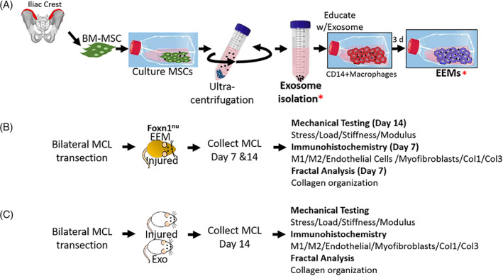
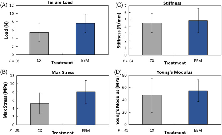
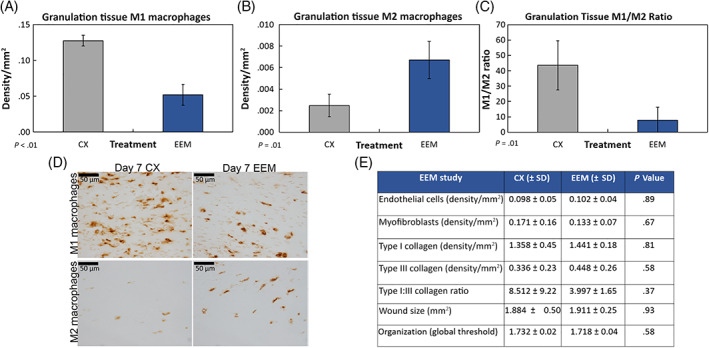
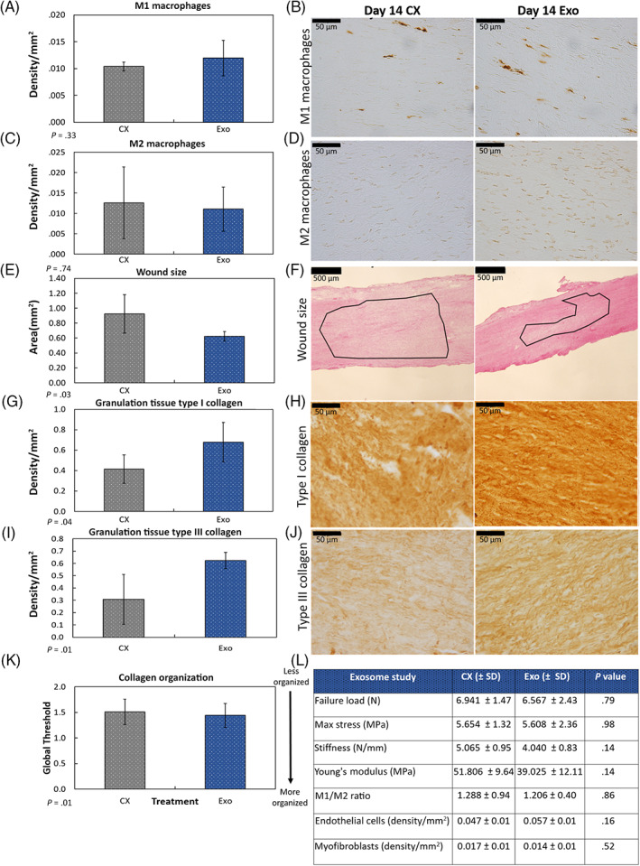
Recently, our group used exosomes from mesenchymal stromal/stem cells (MSCs) to simulate an M2 macrophage phenotype, that is, exosome-educated macrophages (EEMs). These EEMs, when delivered in vivo, accelerated healing in a mouse Achilles tendon injury model. For the current study, we first tested the ability of EEMs to reproduce the beneficial healing effects in a different rodent model, that is, a rat medial collateral ligament (MCL) injury model. We hypothesized that treatment with EEMs would reduce inflammation and accelerate ligament healing, similar to our previous tendon results. Second, because of the translational advantages of a cell-free therapy, exosomes alone were also examined to promote MCL healing. We hypothesized that MSC-derived exosomes could also alter ligament healing to reduce scar formation. Similar to our previous Achilles tendon results, EEMs improved mechanical properties in the healing ligament and reduced inflammation, as indicated via a decreased endogenous M1/M2 macrophage ratio. We also showed that exosomes improved ligament remodeling as indicated by changes in collagen production and organization, and reduced scar formation but without improved mechanical behavior in healing tissue. Overall, our findings suggest EEMs and MSC-derived exosomes improve healing but via different mechanisms. EEMs and exosomes each have attractive characteristics as therapeutics. EEMs as a cell therapy are terminally differentiated and will not proliferate or differentiate. Alternatively, exosome therapy can be used as a cell free, shelf-stable therapeutic to deliver biologically active components. Results herein further support using EEMs and/or exosomes to improve ligament healing by modulating inflammation and promoting more advantageous tissue remodeling.

Keywords: exosomes; ligament healing; macrophages; mesenchymal stromal cells.


Conflict of interest statement

C.S.C. declared employment/leadership position and stock ownership in Dianomi Therapeutics. A.M.S. declared consultant/advisory role with Stryker Endoscopy. M.A.H. declared research funding from OREF Grant Funding. R.V. declared patent holder for EEM technology filed by Wisconsin Alumni Research Foundation. The other authors declared no potential conflicts of interest.

Figures

FIGURE 1
FIGURE 1
Fabrication of exosome‐educated macrophages (EEMs) and experimental design for EEM and exosome studies. A, Bone marrow mesenchymal stromal cells (MSCs) were expanded. Exosomes (Exo) were then isolated from the MSCs via ultracentrifugation. CD14+ monocytes were isolated from human peripheral blood. Monocytes were cultured 7 days, activated to CD14+ macrophages, and then educated with Exo for 3 days, producing EEMs. Two experiments were performed. B, For the first study, Foxn1 nu (nude) rats were subjected to bilateral MCL transection. Immediately after injury, ligaments were treated with EEMs (ipsilateral) or PBS (contralateral). At 7 and 14 days postinjury, MCLs were collected and used for mechanical testing (day 14) or immunohistochemistry (day 7 and day 14). C, For the second study, Wistar rats were subjected to bilateral MCL transection. Rats were treated with either Exo or PBS (serving as the control). At 14 days postinjury, MCLs were collected and used for mechanical testing and immunohistochemistry.* indicates the stage when sample was used for in vivo work
FIGURE 2
FIGURE 2
Mechanical results of the healing medial collateral ligament (MCL) after exosome‐educated macrophages (EEM) treatment. Treatment of the injured MCL with EEMs significantly increased (A) failure load and (B) maximum stress, 14 days postinjury compared to the PBS control (CX). No treatment differences were noted in (C) stiffness or (D) Young's modulus. Data are results of Fisher's LSD post hoc pairwise analysis from 10 MCLs/treatment, (P ≤ .05). Results are expressed as mean ± SD
FIGURE 3
FIGURE 3
Macrophage immunohistochemistry results of the day 7 healing medial collateral ligament (MCL) after treatment with exosome‐educated macrophages (EEMs). Treatment with EEM significantly (A) reduced the number of M1 macrophages, (B) increased the M2 macrophages, and (C) reduced the overall M1/M2 macrophage ratio localized within the granulation tissue compared to the PBS control (CX). Representative images of the (D) M1 macrophages (top) and M2 macrophages (bottom) by the CX (left) and EEM (right) treated MCLs collected 7 days postinjury. E, Table showing results of tested factors that were not significantly different after EEM treatment. Data are considered significantly different (P ≤ .05) based on Fisher's LSD post hoc pairwise analysis from four MCLs/treatment. Values are expressed as mean density/mm2 ± SD. Scale bars = 50 μm
FIGURE 4
FIGURE 4
Immunohistochemistry and histology results by the day 14 healing medial collateral ligament (MCL) after exosome treatment. Treatment of the injured MCL with exosomes (Exo) had no significant effect on the number of (A,B) M1 macrophages, or (C,D) M2 macrophages 14 days postinjury. E,F, In contrast, Exo treatment significantly (E) reduced size of scar tissue compared to the control. F, Representative H&E stained images indicate the reduction in scar size. Outlined region (ie, border between the organized and disorganized collagen fibers) indicates wound area. Exo treatment also significantly increased (G,H) type I and (I,J) type III collagen production within the granulation tissue. K, Exo treatment significantly improved collagen organization. L, Table showing results of tested factors that were not significantly different after Exo treatment. Data are considered significantly different (P < .05) based on Fisher's LSD post hoc pairwise analysis from five MCLs/treatment. Values are expressed as mean area or density/mm2 ± SD

References

    1. Chamberlain CS, Clements AEB, Kink JA, et al. Extracellular vesicle‐educated macrophages promote early Achilles tendon healing. Stem Cells. 2019;37:652‐662. - PMC - PubMed
    1. Kink JA, Forsberg MH, Reshetylo S, et al. Macrophages educated with exosomes from primed mesenchymal stem cells treat acute radiation syndrome by promoting hematopoietic recovery. Biol Blood Marrow Transplant. 2019;25:2124‐2133. - PMC - PubMed
    1. Eslani M, Putra I, Shen X, et al. Cornea‐derived mesenchymal stromal cells therapeutically modulate macrophage immunophenotype and angiogenic function. Stem Cells. 2018;36:775‐784. - PMC - PubMed
    1. Trams EG, Lauter CJ, Salem N Jr, Heine U. Exfoliation of membrane ecto‐enzymes in the form of micro‐vesicles. Biochim Biophys Acta. 1981;645:63‐70. - PubMed
    1. Elahi FM, Farwell DG, Nolta JA, Anderson JD. Preclinical translation of exosomes derived from mesenchymal stem/stromal cells. Stem Cells. 2020;38:15‐21. - PMC - PubMed

Publication types