. 2021 Jul;31(4):e12918.
doi: 10.1111/bpa.12918. Epub 2021 Jan 28.

Intracranial mesenchymal tumor with FET-CREB fusion-A unifying diagnosis for the spectrum of intracranial myxoid mesenchymal tumors and angiomatoid fibrous histiocytoma-like neoplasms

Affiliations

Intracranial mesenchymal tumor with FET-CREB fusion-A unifying diagnosis for the spectrum of intracranial myxoid mesenchymal tumors and angiomatoid fibrous histiocytoma-like neoplasms

Emily A Sloan et al. Brain Pathol. 2021 Jul.

Abstract

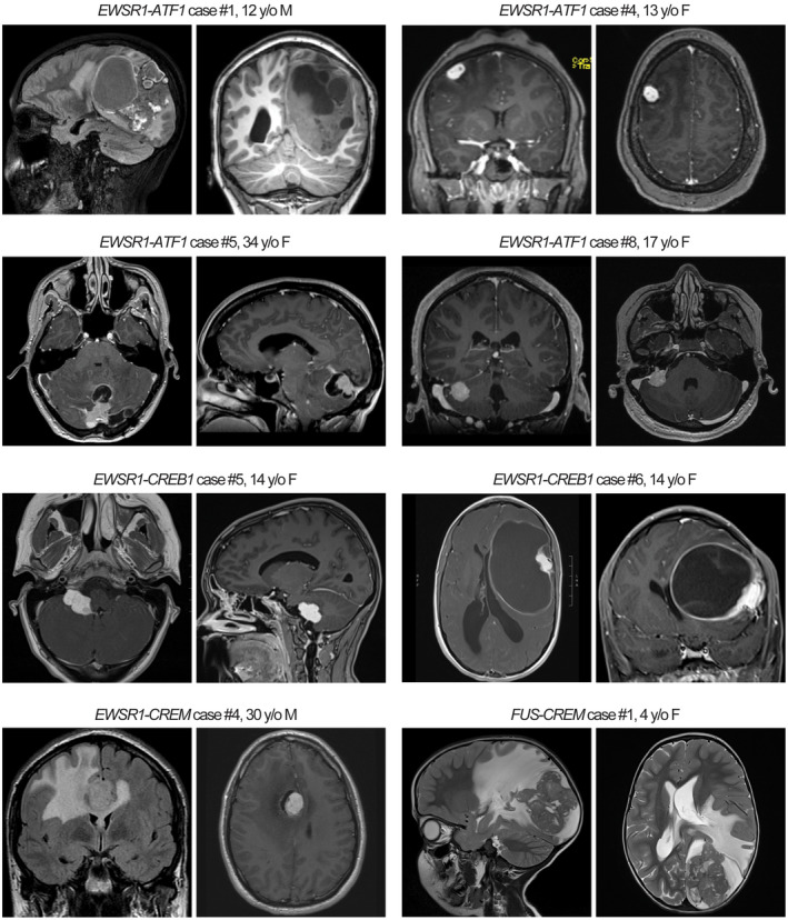
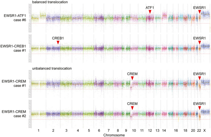
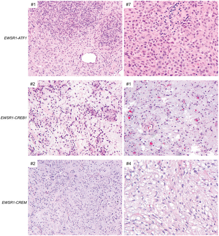
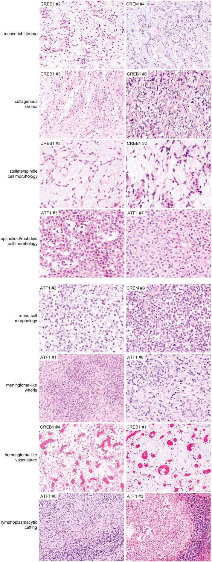
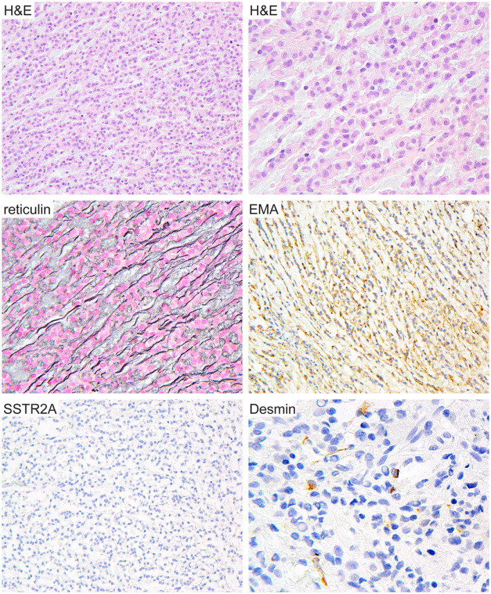
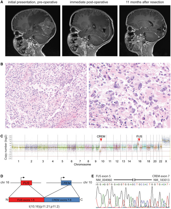
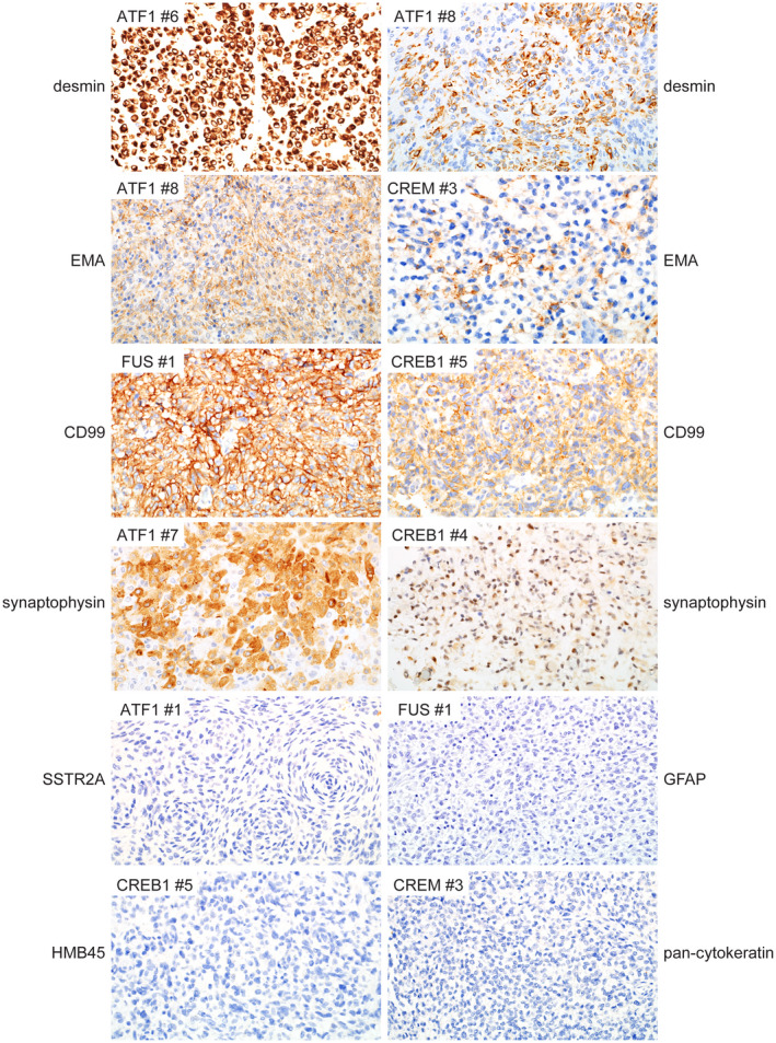
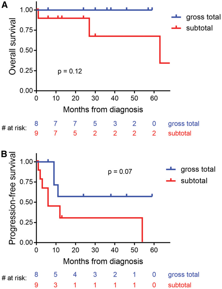
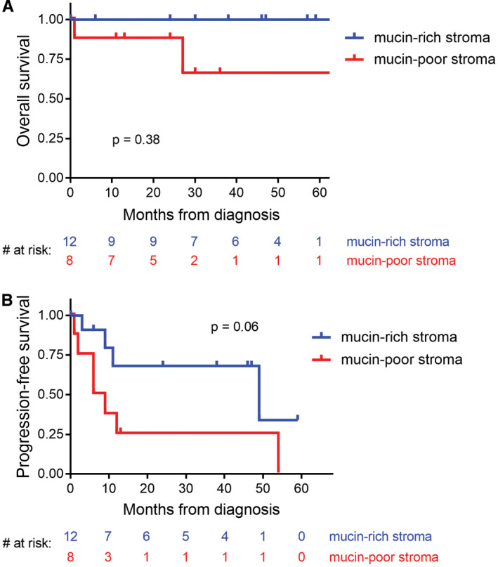
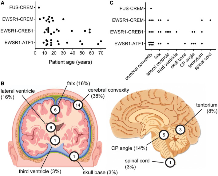
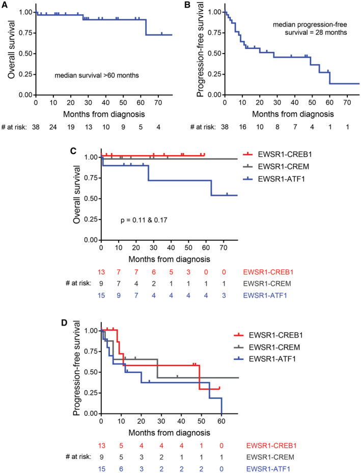
Intracranial mesenchymal tumors with FET-CREB fusions are a recently described group of neoplasms in children and young adults characterized by fusion of a FET family gene (usually EWSR1, but rarely FUS) to a CREB family transcription factor (ATF1, CREB1, or CREM), and have been variously termed intracranial angiomatoid fibrous histiocytoma or intracranial myxoid mesenchymal tumor. The clinical outcomes, histologic features, and genomic landscape are not well defined. Here, we studied 20 patients with intracranial mesenchymal tumors proven to harbor FET-CREB fusion by next-generation sequencing (NGS). The 16 female and four male patients had a median age of 14 years (range 4-70). Tumors were uniformly extra-axial or intraventricular and located at the cerebral convexities (n = 7), falx (2), lateral ventricles (4), tentorium (2), cerebellopontine angle (4), and spinal cord (1). NGS demonstrated that eight tumors harbored EWSR1-ATF1 fusion, seven had EWSR1-CREB1, four had EWSR1-CREM, and one had FUS-CREM. Tumors were uniformly well circumscribed and typically contrast enhancing with solid and cystic growth. Tumors with EWSR1-CREB1 fusions more often featured stellate/spindle cell morphology, mucin-rich stroma, and hemangioma-like vasculature compared to tumors with EWSR1-ATF1 fusions that most often featured sheets of epithelioid cells with mucin-poor collagenous stroma. These tumors demonstrated polyphenotypic immunoprofiles with frequent positivity for desmin, EMA, CD99, MUC4, and synaptophysin, but absence of SSTR2A, myogenin, and HMB45 expression. There was a propensity for local recurrence with a median progression-free survival of 12 months and a median overall survival of greater than 60 months, with three patients succumbing to disease (all with EWSR1-ATF1 fusions). In combination with prior case series, this study provides further insight into intracranial mesenchymal tumors with FET-CREB fusion, which represent a distinct group of CNS tumors encompassing both intracranial myxoid mesenchymal tumor and angiomatoid fibrous histiocytoma-like neoplasms.

Keywords: CREB; EWSR1; angiomatoid fibrous histiocytoma (AFH); brain tumor; intracranial myxoid mesenchymal tumor; molecular neuropathology; sarcoma.


Conflict of interest statement

The authors declare that they have no competing interests.

Figures

FIGURE 1
FIGURE 1
Radiologic features of intracranial mesenchymal tumors with FET‐CREB fusion. These tumors were typically contrast enhancing and often demonstrated both solid and cystic components with extensive peritumoral edema
FIGURE 2
FIGURE 2
Chromosomal copy number analysis demonstrates that EWSR1ATF1 and EWSR1CREB1 fusions typically result from balanced translocations, whereas EWSR1CREM fusions typically result from unbalanced translocations
FIGURE 3
FIGURE 3
Histologic features of intracranial mesenchymal tumors with FET‐CREB fusion. Those tumors with EWSR1ATF1 fusion were most often composed of sheets of epithelioid to rhabdoid cells in a mucin‐poor collagenous stroma. Those tumors with EWSR1CREB1 or EWSR1CREM fusions were most often composed of cords and clusters of stellate to spindle cells in a mucin‐rich stroma
FIGURE 4
FIGURE 4
Specific histologic features recurrently observed in intracranial mesenchymal tumors with FET‐CREB fusion. Example illustrations are shown of the characteristic histologic features recurrently observed across all fusion types (e.g., collagenous stroma, lymphoplasmacytic cuffing) and those enriched in specific fusion types (e.g., epithelioid/rhabdoid cell morphology with EWSR1ATF1 fusion; mucin‐rich stroma, stellate/spindle cell morphology, and hemangioma‐like vasculature with EWSR1CREB1 fusion)
FIGURE 5
FIGURE 5
Intracranial mesenchymal tumor with FET‐CREB fusion can histologically mimic chordoid meningioma. Patient ATF1 #2 is a 9‐year‐old female who presented with an extra‐axial mass along the cerebral convexity of the left frontal lobe. Surgical resection demonstrated a neoplasm composed of cords and clusters of epithelioid cells in a mucin‐rich stroma closely resembling chordoid meningioma. However, the tumor was negative for SSTR2A expression and contained small foci of tumor cells with desmin expression. Genomic interrogation revealed an in‐frame EWSR1ATF1 fusion and absence of mutations involving NF2 and other genes characteristic of meningioma
FIGURE 6
FIGURE 6
Intracranial mesenchymal tumor with FUSCREM fusion. A, A 4‐year‐old girl presented with fevers and headaches and was found to have a large circumscribed and heterogeneously‐enhancing mass in the left occipital region of the brain with significant peritumoral edema. After gross total resection, local tumor recurrence was seen on follow‐up imaging as multiple‐enhancing nodules at the periphery of the prior resection cavity. B, Histologic sections demonstrated a highly cellular neoplasm with both sheet‐like and papillary growth patterns composed of epithelioid to rhabdoid tumor cells with prominent nuclear pleomorphism and atypia. C, Chromosomal copy number plot demonstrates that the FUSCREM fusion was the result of an unbalanced translocation between chromosome 10p11.21 and chromosome 16p11.2. D, Schematic of the FUSCREM gene fusion. E, Sanger chromatogram following reverse‐transcription PCR of the FUSCREM fusion transcript composed of exons 1–5 of FUS linked with exons 7–8 of CREM
FIGURE 7
FIGURE 7
Immunohistochemical features of intracranial mesenchymal tumors with FET‐CREB fusion. These tumors were uniformly positive for desmin expression, which ranged from diffuse strong labeling to cases with focal clusters of positive cells only. Most tumors were also positive for EMA and CD99 expression in a membranous distribution. A subset of tumors demonstrated positivity for synaptophysin that ranged from diffuse strong to more focal and weak staining. These tumors were uniformly negative for SSTR2A, GFAP, and HMB45. Most tumors were negative for cytokeratin expression, with only a single tumor that demonstrated focal positivity for cytokeratin AE1/AE3 (not shown)
FIGURE 8
FIGURE 8
Clinical outcomes for patients with intracranial mesenchymal tumors with FET‐CREB fusion based on extent of surgical resection. A, Kaplan–Meier plot of the overall survival for the 17 patients from this study with known extent of resection. B, Kaplan–Meier plot of progression‐free survival for the 17 patients from this study with known extent of resection. Compared to gross total resection, subtotal resection was associated with increased risk of death and tumor recurrence (although neither was statistically significant)
FIGURE 9
FIGURE 9
Clinical outcomes for patients with intracranial mesenchymal tumors with FET‐CREB fusion based on mucin content of the stroma. A and B, Kaplan–Meier plot of overall survival (A) and progression‐free survival (B) for the 20 patients from this study stratified by mucin‐rich versus mucin‐poor stroma
FIGURE 10
FIGURE 10
Anatomic location and age at diagnosis for intracranial mesenchymal tumors with FET‐CREB fusion from this study combined with all previously reported cases in the literature (see Table 4). A, Plot of patient age at diagnosis versus gene fusion. B, Diagram of tumor locations. C. Plot of tumor location versus gene fusion
FIGURE 11
FIGURE 11
Clinical outcomes for patients with intracranial mesenchymal tumors with FET‐CREB fusion from this study combined with all previously reported cases in the literature (see Table 4). A, Kaplan–Meier plot of overall survival demonstrates a median overall survival >60 months. B, Kaplan–Meier plot of progression‐free survival demonstrates that a majority of patients experience disease recurrence with a median interval of 28 months. C, Kaplan–Meier plot of overall survival stratified by fusion type. While no statistically significant difference in overall survival based on fusion type is observed, three patients with tumors containing EWSR1ATF1 fusion succumbed to disease, while all patients with EWSR1CREB1 or EWSR1CREM fusion remained alive at time of last clinical follow‐up. D, Kaplan–Meier plot of progression‐free survival stratified by fusion type. No significant difference was observed based on fusion type (median recurrence‐free survival: EWSR1ATF1, 16 months; EWRS1CREB1, 49 months; EWSR1CREM, 28 months)

References

    1. Bale TA, Oviedo A, Kozakewich H, Giannini C, Davineni PK, Ligon K, et al. Intracranial myxoid mesenchymal tumors with EWSR1‐CREB family gene fusions: myxoid variant of angiomatoid fibrous histiocytoma or novel entity? Brain Pathol. 2018;28:183–91. - PMC - PubMed
    1. Dunham C, Hussong J, Seiff M, Pfeifer J, Perry A. Primary intracerebral angiomatoid fibrous histiocytoma: report of a case with a t(12;22)(q13;q12) causing type 1 fusion of the EWS and ATF‐1 genes. Am J Surg Pathol. 2008;32:478–84. - PubMed
    1. Kao YC, Sung YS, Zhang L, Chen CL, Vaiyapuri S, Rosenblum MK, et al. EWSR1 fusions with CREB family transcription factors define a novel myxoid mesenchymal tumor with predilection for intracranial location. Am J Surg Pathol. 2017;41:482–90. - PMC - PubMed
    1. Konstantinidis A, Cheesman E, O'Sullivan J, Pavaine J, Avula S, Pizer B, et al. Intracranial angiomatoid fibrous histiocytoma with EWSR1‐CREB family fusions: a report of two pediatric cases. World Neurosurg. 2019;126:113–9. - PubMed
    1. Antonescu CR, Dal Cin P, Nafa K, Teot LA, Surti U, Fletcher CD, et al. EWSR1‐CREB1 is the predominant gene fusion in angiomatoid fibrous histiocytoma. Genes Chromosomes Cancer. 2007;46:1051–60. - PubMed

Publication types

MeSH terms

Substances

Supplementary concepts