

Residual Structure of Unfolded Ubiquitin as Revealed by Hydrogen/Deuterium-Exchange 2D NMR
- PMID: 33142107
- PMCID: PMC7732725
- DOI: 10.1016/j.bpj.2020.10.003
Residual Structure of Unfolded Ubiquitin as Revealed by Hydrogen/Deuterium-Exchange 2D NMR
Abstract
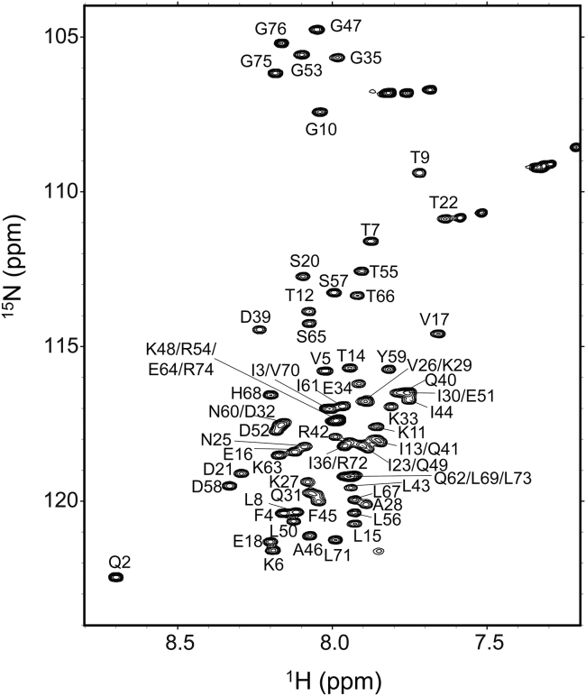
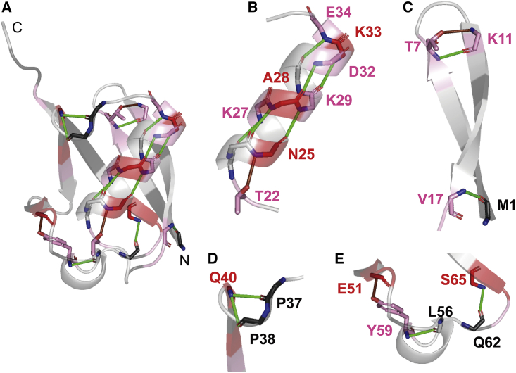
The characterization of residual structures persistent in unfolded proteins in concentrated denaturant solution is currently an important issue in studies of protein folding because the residual structure present, if any, in the unfolded state may form a folding initiation site and guide the subsequent folding reactions. Here, we studied the hydrogen/deuterium (H/D)-exchange behavior of unfolded human ubiquitin in 6 M guanidinium chloride. We employed a dimethylsulfoxide (DMSO)-quenched H/D-exchange NMR technique with the use of spin desalting columns, which allowed us to perform a quick medium exchange from 6 M guanidinium chloride to a quenching DMSO solution. Based on the backbone resonance assignment of ubiquitin in the DMSO solution, we successfully investigated the H/D-exchange kinetics of 60 identified peptide amide groups in the ubiquitin sequence. Although a majority of these amide groups were not protected, certain amide groups involved in a middle helix (residues 23-34) and an N-terminal β-hairpin (residues 2-16) were significantly protected with a protection factor of 2.1-4.2, indicating that there were residual structures in unfolded ubiquitin and that these amide groups were more than 52% hydrogen bonded in the residual structures. We show that the hydrogen-bonded residual structures in the α-helix and the β-hairpin are formed even in 6 M guanidinium chloride, suggesting that these residual structures may function as a folding initiation site to guide the subsequent folding reactions of ubiquitin.
Copyright © 2020 Biophysical Society. Published by Elsevier Inc. All rights reserved.
Figures


Similar articles
-
DMSO-Quenched H/D-Exchange 2D NMR Spectroscopy and Its Applications in Protein Science.Molecules. 2022 Jun 10;27(12):3748. doi: 10.3390/molecules27123748. Molecules. 2022. PMID: 35744871 Free PMC article. Review.
-
The B domain of protein A retains residual structures in 6 M guanidinium chloride as revealed by hydrogen/deuterium-exchange NMR spectroscopy.Protein Sci. 2023 Mar;32(3):e4569. doi: 10.1002/pro.4569. Protein Sci. 2023. PMID: 36659853 Free PMC article.
-
The use of spin desalting columns in DMSO-quenched H/D-exchange NMR experiments.Protein Sci. 2013 Apr;22(4):486-91. doi: 10.1002/pro.2221. Epub 2013 Feb 11. Protein Sci. 2013. PMID: 23339068 Free PMC article.
-
Investigation of ribonuclease T1 folding intermediates by hydrogen-deuterium amide exchange-two-dimensional NMR spectroscopy.Biochemistry. 1993 Jun 22;32(24):6152-6. doi: 10.1021/bi00075a006. Biochemistry. 1993. PMID: 8512924
-
Measuring the hydrogen/deuterium exchange of proteins at high spatial resolution by mass spectrometry: overcoming gas-phase hydrogen/deuterium scrambling.Acc Chem Res. 2014 Oct 21;47(10):3018-27. doi: 10.1021/ar500194w. Epub 2014 Aug 29. Acc Chem Res. 2014. PMID: 25171396 Review.
Cited by
-
DMSO-Quenched H/D-Exchange 2D NMR Spectroscopy and Its Applications in Protein Science.Molecules. 2022 Jun 10;27(12):3748. doi: 10.3390/molecules27123748. Molecules. 2022. PMID: 35744871 Free PMC article. Review.
-
The B domain of protein A retains residual structures in 6 M guanidinium chloride as revealed by hydrogen/deuterium-exchange NMR spectroscopy.Protein Sci. 2023 Mar;32(3):e4569. doi: 10.1002/pro.4569. Protein Sci. 2023. PMID: 36659853 Free PMC article.
-
Recent Advances in NMR Protein Structure Prediction with ROSETTA.Int J Mol Sci. 2023 Apr 25;24(9):7835. doi: 10.3390/ijms24097835. Int J Mol Sci. 2023. PMID: 37175539 Free PMC article. Review.
References
-
- Tanford C. Protein denaturation. Adv. Protein Chem. 1968;23:121–282. - PubMed
-
- Tanford C. Protein denaturation. C. Theoretical models for the mechanism of denaturation. Adv. Protein Chem. 1970;24:1–95. - PubMed
-
- Neri D., Billeter M., Wüthrich K. NMR determination of residual structure in a urea-denatured protein, the 434-repressor. Science. 1992;257:1559–1563. - PubMed
-
- Schwalbe H., Fiebig K.M., Dobson C.M. Structural and dynamical properties of a denatured protein. Heteronuclear 3D NMR experiments and theoretical simulations of lysozyme in 8 M urea. Biochemistry. 1997;36:8977–8991. - PubMed
-
- Klein-Seetharaman J., Oikawa M., Schwalbe H. Long-range interactions within a nonnative protein. Science. 2002;295:1719–1722. - PubMed
Publication types
MeSH terms
Substances
LinkOut - more resources
Full Text Sources
