. 2020 Nov;99(11):6084-6093.
doi: 10.1016/j.psj.2020.07.024. Epub 2020 Aug 8.

Heat stress decreases egg production of laying hens by inducing apoptosis of follicular cells via activating the FasL/Fas and TNF-α systems

Affiliations

Heat stress decreases egg production of laying hens by inducing apoptosis of follicular cells via activating the FasL/Fas and TNF-α systems

Gui-Ming Li et al. Poult Sci. 2020 Nov.

Abstract

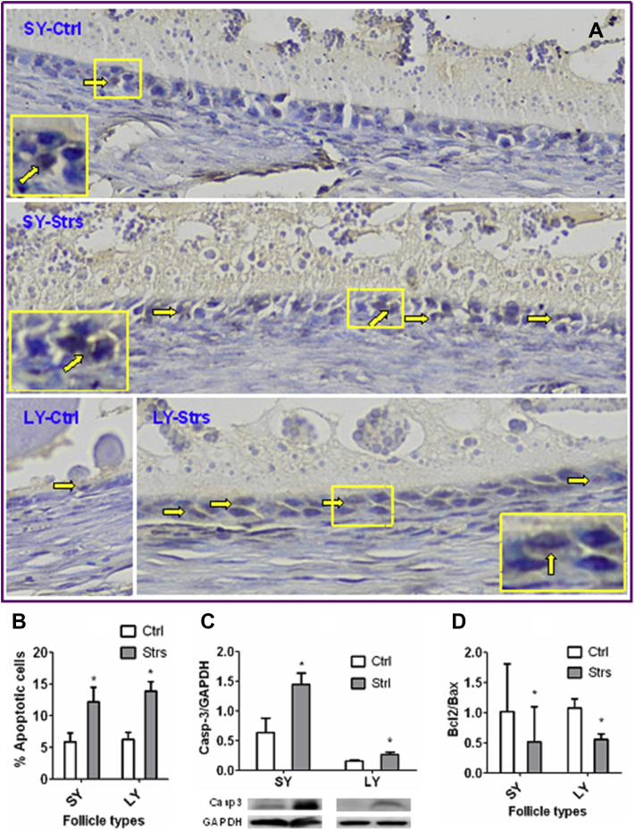
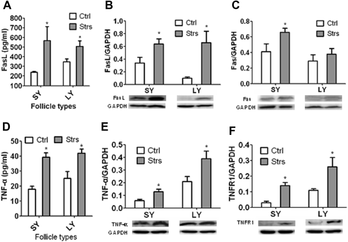
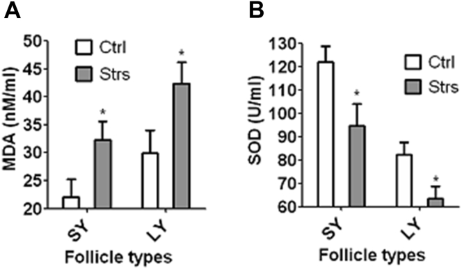
Heat stress (HS) causes significant economic losses in the poultry industry every year. However, the mechanisms for the adverse effects of HS on avian follicular development are largely unknown. The aim of this study was to test whether HS induces apoptosis of follicular cells and impairs egg production by activating the FasL/Fas and tumor necrosis factor (TNF)-α systems. To this end, Hy-Line Brown laying hens, at 32 wk of age, were either exposed to HS of 35°C to 37°C or maintained at 24°C to 26°C (control) for 5 D. At the end of the HS period, follicle numbers, apoptosis, FasL/Fas and TNF-α activation, oxidative stress, and hormone secretion were examined in ovarian follicles. Egg production was observed daily during both the stressed (day S1-S5) and the poststress recovery (day R1-R15) periods. The results demonstrated that HS on hens significantly 1) decreased laying rates from day S3 to R6; 2) reduced numbers of large yellow and hierarchical follicles; 3) triggered apoptosis while increasing the expression of FasL, Fas, TNF-α, and TNF-receptor 1 in small and large yellow follicles; and 4) increased levels of oxidative stress, corticotrophin-releasing hormone, and corticosterone while decreasing the estradiol/progesterone ratio in follicular fluid in small and large yellow follicles. Taken together, the results suggested that hen HS impaired egg production by reducing the number of follicles through inducing apoptosis and that it triggered apoptosis in follicular cells by activating the FasL/Fas and TNF-α systems.

Keywords: FasL/Fas signaling; TNF-α signaling; follicular cell apoptosis; heat stress; laying hen.


Figures

Figure 1
Figure 1
Effects of heat stress of laying hens on laying rates and follicle development. Graph (A) shows percentages of laying/total hens on each heat stressed (S) or poststress recovering (R) day in control (Ctrl) or stressed (Strs) hens. In this experiment, 120 layers were randomly divided into stressed and control groups each containing 60 hens. Egg production was observed daily during both the stress period (day S1 to S5) and the poststress recovery period (day R1 to R15). On each day, each treatment was repeated 20 times with each replicate containing 3 hens. Graph (B) compares numbers of large white (LW), small yellow (SY), large yellow (LY), and hierarchical (Hie) follicles between Ctrl and Strs hens on day 5 of heat stress. Each treatment was repeated 10 times with each replicate containing one ovary. ∗ indicates significant difference (P < 0.05) from control values within d or follicle types. Panel (C) shows the estimated ovulation times for LW, SY, LY, and hierarchical follicles from the onset of heat stress exposure. From the onset of heat stress, it took 11 D for the LW follicles of 3 to 5 mm in diameter, 8 D for the SY follicles of 5 to 8 mm in diameter, 6 D– for the LY follicles of 8 to 12 mm in diameter, and about 3 to 5 D for the F3 to F5 hierarchical follicles of >12 mm in diameter to grow and mature before ovulation (Δ).
Figure 2
Figure 2
Effects of heat stress of laying hens on apoptosis of follicles. Panel (A) shows images of follicular wall sections after TUNEL staining of SY or LY follicles from control (Ctrl) or stressed (Strs) hens. In the images, while the TUNEL-positive nuclei of mural granulosa cells (MGC) were stained brown (arrows), the TUNEL-negative nuclei appear blue. Boxed regions in the image are shown as 2-fold enlarged insets to highlight the TUNEL-positive brown nuclei. Graphs (B), (C), and (D) compare percentages of apoptotic cells (TUNEL-positive), levels of active caspase-3 (Caspase-3/GAPDH ratio, Western blotting), and Bcl2/Bax ratio (RT-PCR results), respectively, in MGC of SY or LY follicles between Ctrl and Strs hens on day 5 of heat stress. In graph (B), each treatment was repeated 6 times with each replicate containing 1 hen contributing 2 SY or 1 or 2 LY follicles. In graph (C), each treatment was repeated 5 times with each replicate containing 2 hens each contributing 2 SY follicles or 6 times with each replicate containing 2 hens each contributing 1–3 LY follicles. In graph (D), each treatment was repeated 8 times with each replicate containing 1 SY or LY follicle from 1 hen. ∗ indicates significant difference (P < 0.05) from control values within follicle types. Abbreviations: LY, large yellow; SY, small yellow; TUNEL, Terminal deoxynucleotidyl transferase dUTP Nick-End Labeling.
Figure 3
Figure 3
Effects of heat stress of laying hens on FasL/Fas and TNF-α/TNFR1 expression in ovarian follicles. Graphs (A) and (D) show ELISA results comparing FasL and TNF-α levels, respectively, in follicular fluid from SY or LY follicles between Ctrl and Strs hens on day 5 of heat stress. Each treatment was repeated 6 times and each replicate contained 1 hen contributing pooled follicular fluid from 3 SY or 1–3 LY follicles. Graphs (B)/(C) and (E)/(F) show Western blotting results comparing levels of FasL/Fas (Fasl or Fas/GAPDH ratio) and TNF-α/TNFR1 (TNF-α or TNFR1/GAPDH ratio), respectively, in MGC from SY or LY follicles between Ctrl and Strs hens. Each treatment was repeated 7 times with each replicate including MGC from 2 hens each contributing 2 SY follicles, or repeated 8 times with each replicates including 2 hens each contributing 1–3 LY follicles. ∗ indicates significant difference (P < 0.05) from control values within follicle types. Abbreviations: Ctrl, control hens; LY, large yellow; Strs, stressed hens; SY, small yellow; TNF, tumor necrosis factor.
Figure 4
Figure 4
Effects of heat stress of laying hens on redox status of SY and LY follicles. Graphs (A) and (B) show malondialdehyde (MDA) and superoxide dismutase (SOD) levels, respectively, in follicular fluid from SY and LY follicles of control (Ctrl) or stressed (Strs) hens on day 5 of heat stress. In graph (A), each treatment was repeated 8 times with each replicate containing 1 hen contributing pooled follicular fluid from 3 SY follicles or 1–3 LY follicles. In graph (B), each treatment was repeated 5 times with each replicate containing 1 hen contributing pooled follicular fluid from 3 SY or 1-3 LY follicles. ∗ indicates significant difference (P < 0.05) from control values within follicle types. Abbreviations: LY, large yellow; SY, small yellow.
Figure 5
Figure 5
Effects of heat stress of laying hens on hormone levels in serum and follicular fluid. Hormone concentrations were measured by ELISA. Graphs (A), (B), and (C) compare E2 and P4 concentrations and the E2/P4 ratio, respectively, in serum or follicular fluid from SY or LY follicles between Ctrl and Strs hens on day 5 of heat stress. Each treatment was repeated 5 times with each replicate containing 1 serum sample from 1 hen, or repeated 9 times with each replicate containing follicular fluid from 1 follicle from 1 hen. Graphs (D) and (E) compare levels of CRH and corticosterone, respectively, in serum or follicular fluid from SY or LY follicles between Ctrl and Strs hens. Each treatment was repeated 6 times with each replicate containing 1 hen providing 1 serum sample or follicular fluid from 3 SY or 1–3 LY follicles. ∗ indicates significant difference (P < 0.05) from control values within serum or follicle types. Abbreviations: CRH, corticotrophin-releasing hormone; Ctrl, control hens; E2, estradiol; LY, large yellow; P4, progesterone; Strs, stressed hens; SY, small yellow.

References

    1. Alhenaky A., Abdelqader A., Abuajamieh M., Al-Fataftah A.R. The effect of heat stress on intestinal integrity and Salmonella invasion in broiler birds. J. Therm. Biol. 2017;70(Pt B):9–14. - PubMed
    1. Al-Saffar A.A., Rose S.P. Ambient temperature and the egg laying characteristics of the laying fowl. World’s Poult. Sci. J. 2002;58:317–331.
    1. Cramer T., Kisliouk T., Yeshurun S., Meiri N. The balance between stress resilience and vulnerability is regulated by corticotropin-releasing hormone during the critical postnatal period for sensory development. Dev. Neurobiol. 2015;75:842–853. - PubMed
    1. Cui Y., Wu G., Wang Z., Huang F., Ning Z., Chu L., Yang S., Lv Q., Hu J. Effects of Taurine on broiler aortic endothelial apoptosis induced by heat stress. Adv. Exp. Med. Biol. 2019;1155:391–406. - PubMed
    1. Deng W., Dong X.F., Tong J.M., Zhang Q. The probiotic Bacillus licheniformis ameliorates heat stress-induced impairment of egg production, gut morphology, and intestinal mucosal immunity in laying hens. Poult. Sci. 2012;91:575–582. - PubMed