TMS Correlates of Pyramidal Tract Signs and Clinical Motor Status in Patients with Cervical Spondylotic Myelopathy
- PMID: 33142762
- PMCID: PMC7692772
- DOI: 10.3390/brainsci10110806
TMS Correlates of Pyramidal Tract Signs and Clinical Motor Status in Patients with Cervical Spondylotic Myelopathy
Abstract
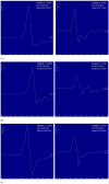
Background: While the association between motor-evoked potential (MEP) abnormalities and motor deficit is well established, few studies have reported the correlation between MEPs and signs of pyramidal tract dysfunction without motor weakness. We assessed MEPs in patients with pyramidal signs, including motor deficits, compared to patients with pyramidal signs but without weakness.
Methods: Forty-three patients with cervical spondylotic myelopathy (CSM) were dichotomized into 21 with pyramidal signs including motor deficit (Group 1) and 22 with pyramidal signs and normal strength (Group 2), and both groups were compared to 33 healthy controls (Group 0). MEPs were bilaterally recorded from the first dorsal interosseous and tibialis anterior muscle. The central motor conduction time (CMCT) was estimated as the difference between MEP latency and peripheral latency by magnetic stimulation. Peak-to-peak MEP amplitude and right-to-left differences were also measured.
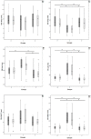
Results: Participants were age-, sex-, and height-matched. MEP latency in four limbs and CMCT in the lower limbs were prolonged, and MEP amplitude in the lower limbs decreased in Group 1 compared to the others. Unlike motor deficit, pyramidal signs were not associated with MEP measures, even when considering age, sex, and height as confounding factors.
Conclusions: In CSM, isolated pyramidal signs may not be associated, at this stage, with MEP changes.
Keywords: cervical spondylotic myelopathy; clinical neuroscience; corticospinal conduction; degenerative cervical myelopathy; motor status; motor-evoked potentials; pyramidal signs; transcranial magnetic stimulation.
Conflict of interest statement
The authors declare no conflict of interest.
Figures


Similar articles
-
Electrophysiological assessments of the motor pathway in diabetic patients with compressive cervical myelopathy.J Neurosurg Spine. 2015 Dec;23(6):707-14. doi: 10.3171/2015.3.SPINE141060. Epub 2015 Sep 4. J Neurosurg Spine. 2015. PMID: 26340381
-
The role of electrophysiology in the diagnosis and management of cervical spondylotic myelopathy.Ann Acad Med Singap. 2007 Nov;36(11):886-93. Ann Acad Med Singap. 2007. PMID: 18071594
-
Sex-specific reference values for total, central, and peripheral latency of motor evoked potentials from a large cohort.Front Hum Neurosci. 2023 Jun 9;17:1152204. doi: 10.3389/fnhum.2023.1152204. eCollection 2023. Front Hum Neurosci. 2023. PMID: 37362949 Free PMC article.
-
Central motor conduction time.Handb Clin Neurol. 2013;116:375-86. doi: 10.1016/B978-0-444-53497-2.00031-0. Handb Clin Neurol. 2013. PMID: 24112910 Review.
-
Pyramidal weakness: Is it time to retire the term?Clin Anat. 2021 Apr;34(3):478-482. doi: 10.1002/ca.23715. Epub 2020 Dec 31. Clin Anat. 2021. PMID: 33347647 Review.
Cited by
-
Normal parameters for diagnostic transcranial magnetic stimulation using a parabolic coil with biphasic pulse stimulation.BMC Neurol. 2022 Dec 31;22(1):510. doi: 10.1186/s12883-022-02977-8. BMC Neurol. 2022. PMID: 36585660 Free PMC article.
-
Challenging the Pleiotropic Effects of Repetitive Transcranial Magnetic Stimulation in Geriatric Depression: A Multimodal Case Series Study.Biomedicines. 2023 Mar 21;11(3):958. doi: 10.3390/biomedicines11030958. Biomedicines. 2023. PMID: 36979937 Free PMC article.
-
Degenerative cervical myelopathy delays responses to lateral balance perturbations regardless of predictability.J Neurophysiol. 2022 Mar 1;127(3):673-688. doi: 10.1152/jn.00159.2021. Epub 2022 Jan 26. J Neurophysiol. 2022. PMID: 35080466 Free PMC article.
-
People with degenerative cervical myelopathy have impaired reactive balance during walking.Gait Posture. 2024 Mar;109:303-310. doi: 10.1016/j.gaitpost.2024.02.014. Epub 2024 Feb 22. Gait Posture. 2024. PMID: 38412683 Free PMC article.
-
Sex differences in mild vascular cognitive impairment: A multimodal transcranial magnetic stimulation study.PLoS One. 2023 Mar 3;18(3):e0282751. doi: 10.1371/journal.pone.0282751. eCollection 2023. PLoS One. 2023. PMID: 36867595 Free PMC article.
References
-
- Rossini P.M., Burke D., Chen R., Cohen L.G., Daskalakis Z., Di Iorio R., Di Lazzaro V., Ferreri F., Fitzgerald P.B., George M.S., et al. Non-invasive electrical and magnetic stimulation of the brain, spinal cord, roots and peripheral nerves: Basic principles and procedures for routine clinical and research application. An updated report from an I.F.C.N. Committee. Clin. Neurophysiol. Off. J. Int. Fed. Clin. Neurophysiol. 2015;126:1071–1107. doi: 10.1016/j.clinph.2015.02.001. - DOI - PMC - PubMed
-
- Lanza G., Bella R., Giuffrida S., Cantone M., Pennisi G., Spampinato C., Giordano D., Malaguarnera G., Raggi A., Pennisi M. Preserved transcallosal inhibition to transcranial magnetic stimulation in nondemented elderly patients with leukoaraiosis. BioMed Res. Int. 2013;2013:351680. doi: 10.1155/2013/351680. - DOI - PMC - PubMed
LinkOut - more resources
Full Text Sources

