Review
. 2020 Oct 31;10(11):1503.
doi: 10.3390/biom10111503.

BRCA1-A and BRISC: Multifunctional Molecular Machines for Ubiquitin Signaling

Affiliations
Review

BRCA1-A and BRISC: Multifunctional Molecular Machines for Ubiquitin Signaling

Julius Rabl. Biomolecules. .

Abstract

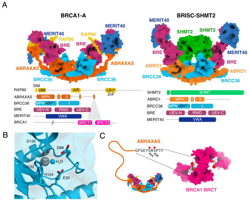
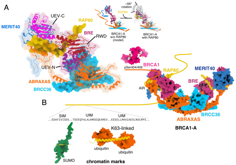
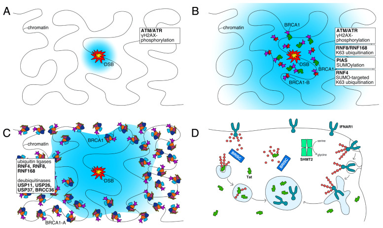
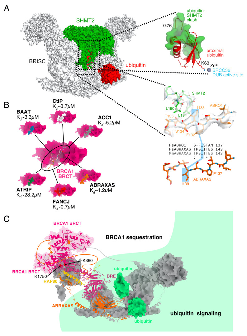
The K63-linkage specific deubiquitinase BRCC36 forms the core of two multi-subunit deubiquitination complexes: BRCA1-A and BRISC. BRCA1-A is recruited to DNA repair foci, edits ubiquitin signals on chromatin, and sequesters BRCA1 away from the site of damage, suppressing homologous recombination by limiting resection. BRISC forms a complex with metabolic enzyme SHMT2 and regulates the immune response, mitosis, and hematopoiesis. Almost two decades of research have revealed how BRCA1-A and BRISC use the same core of subunits to perform very distinct biological tasks.

Keywords: BRCA1; BRCC36; DNA repair; RAP80; SHMT2; SUMO; deubiquitination; immune regulation; ubiquitin.


Conflict of interest statement

The author declares no conflict of interest.

Figures

Figure 1
Figure 1
(A) Architecture of BRCA1-A and BRISC-SHMT2α complex. Schematic representation of protein subunits and binding partners (BRCA1-A and BRISC-SHMT2α structure: [25]). (B) Detailed view of the active site of BRCC36. The side chains of catalytic residues E33, D88, H122, H124, and D135 are shown. The zinc atom at the active center is shown in silver. A catalytic water molecule is shown as a blue sphere (BRCA1-A structure: [25]). (C) The BRCA1 BRCT dimer binds to the phosphorylated C-terminus of ABRAXAS. The flexible C-terminus of ABRAXAS is depicted schematically and the conserved BRCT binding sequence of ABRAXAS is shown. The phosphorylation sites at S404 and S406 are indicated with red dots. The BRCA1 BRCT domain dimer bound to the phosphorylated ABRAXAS C-terminus is shown in surface representation with the BRCA1 shown in pink and the ABRAXAS C-terminal tail in white with phosphorylation sites shown in red (BRCA1 BRCT structure: [30], BRCA1-A structure: [25]).
Figure 2
Figure 2
(A) Integration of RAP80 into the BRCA1-A complex. RAP80 and BRCC36 are shown as surface representation, ABRAXAS, BRE and MERIT40 are shown as cartoon. Integration of RAP80 into BRCA1-A induces a substantial conformation change in BRE, which results in a rotation of ∼56 towards the coiled coils at the base of the complex (BRCA1-A and BRISC-SHMT2α structure: [25]). (B) Schematic representation of BRCA1-A binding to chromatin marks. The SIM and twin UIM domains at the N-terminus of RAP80 are tethered by a long flexible linker to BRCA1-A. BRCA1 binds to the C-terminus of ABRAXAS (BRCA1-A structure: [25], BRCA1 BRCT domain with bound ABRAXAS phosphopeptide structure: [30], RAP80 SIM-bound ubiquitin structure: [41], RAP80 UIM-bound K63-linked ubiquitin structure: [42]).
Figure 3
Figure 3
Schematic representation of DNA double-strand break repair associated signaling events (AC) and interferon and Tat degradation control by BRISC (D). (A) A DNA double-strand break (red, center) has been detected, ARM/ATR marks chromatin surrounding the site of damage (blue). (B) Signaling by ubiquitin ligases, sumo ligases, and SUMO-targeted ubiquitin ligases marks chromatin surrounding the site of damage (blue) while BRCA1 is recruited to the site of damage where it engages in resection-promoting complexes. (C) DUBs including BRCC36 edit the chromatin marks (blue) and limit their spread. BRCA1 is sequestered to the periphery of the DNA repair focus and withdrawn from the site of damage. (D) BRISC degrades K63-linked chains that serve as lysosomal degradation signals from membrane receptor IFNAR1 and soluble viral protein Tat, increasing their stability and concentration.
Figure 4
Figure 4
(A) Inactivation of BRISC by SHMT2α binding. A cryo-EM map of BRISC-SHMT2α complex is shown with a di-ubiquitin fit into the active site using the structure of AMSH-LP with bound di-ubiquitin [80]. The clash between SHMT2 and the proximal ubiquitin is shown in a zoom-out. BRCA1-A complex does not bind SHMT2, because residue I133 of Abro1, which is essential for the interaction, is conserved, but flipped by 180 due to a proline insertion at position P137. The cryo-EM map of BRISC-SHMT2α is shown in white while the side chains of SHMT2 and ABRO1 are shown in green and light orange, respectively. The structure of ABRAXAS that corresponds to the part of ABRO1 shown is depicted below in orange (BRCA1-A and BRISC-SHMT2α structure: [25]). (B) ACC1, ATRIP, BAAT, CtIP, and FANCJ compete with the phosphorylated C-terminus of ABRAXAS for the same binding site on BRCA1. Binding affinities measured for the phosphorylated peptide only are in the micromolar range. The entire BRCA1-A complex however binds with nanomolar affinity (ABRAXAS-BRCA1 complex structure: [30], ACC1-BRCA1 complex structure: [83], ATRIP-BRCA1 complex structure: [84], BAAT-BRCA1 complex structure: [84], CtIP-BRCA1 complex structure: [85], FANCJ-BRCA1 complex structure: [86]). (C) Crosslinking data suggests that the BRCA1 BRCT domain is localized at the edge of the arc, distal from the active sites of the complex. This model explains why sequestration of BRCA1 by BRCA1-A does not affect the DUB activity of the complex. BRCA1-A can be considered to be divided into a ubiquitin signaling zone (green) and a BRCA1 sequestration zone that are functionally independent (BRCA1-A structure: [25], AMSH-LP-substrate complex structure used for modelling ubiquitin at the active site of BRCA1-A: [80], structure of BRCA1 BRCT dimer with bound ABRAXAS C-terminal phosphorylated peptide: [30]).

Similar articles

Cited by

References

    1. Komander D., Rape M. The Ubiquitin Code. Annu. Rev. Biochem. 2012;81:203–229. doi: 10.1146/annurev-biochem-060310-170328. - DOI - PubMed
    1. Kauko A., Lehto K. Eukaryote specific folds: Part of the whole. Proteins Struct. Funct. Bioinform. 2018;86:868–881. doi: 10.1002/prot.25517. - DOI - PubMed
    1. Delley C.L., Müller A.U., Ziemski M., Weber-Ban E. Prokaryotic Ubiquitin-Like Protein and Its Ligase/Deligase Enyzmes. J. Mol. Biol. 2017;429:3486–3499. doi: 10.1016/j.jmb.2017.04.020. - DOI - PubMed
    1. Polge C., Uttenweiler-Joseph S., Leulmi R., Heng A.E., Burlet-Schiltz O., Attaix D., Taillandier D. Deciphering the ubiquitin proteome: Limits and advantages of high throughput global affinity purification-mass spectrometry approaches. Int. J. Biochem. Cell Biol. 2013;45:2136–2146. doi: 10.1016/j.biocel.2013.05.031. - DOI - PubMed
    1. Hospenthal M.K., Freund S.M.V., Komander D. Assembly, analysis and architecture of atypical ubiquitin chains. Nat. Struct. Mol. Biol. 2013;20:555–565. doi: 10.1038/nsmb.2547. - DOI - PMC - PubMed

LinkOut - more resources