. 2020 Oct 31;9(11):2390.
doi: 10.3390/cells9112390.

Dysbindin deficiency Alters Cardiac BLOC-1 Complex and Myozap Levels in Mice

Affiliations

Dysbindin deficiency Alters Cardiac BLOC-1 Complex and Myozap Levels in Mice

Ankush Borlepawar et al. Cells. .

Abstract

Dysbindin, a schizophrenia susceptibility marker and an essential constituent of BLOC-1 (biogenesis of lysosome-related organelles complex-1), has recently been associated with cardiomyocyte hypertrophy through the activation of Myozap-RhoA-mediated SRF signaling. We employed sandy mice (Dtnbp1_KO), which completely lack Dysbindin protein because of a spontaneous deletion of introns 5-7 of the Dtnbp1 gene, for pathophysiological characterization of the heart. Unlike in vitro, the loss-of-function of Dysbindin did not attenuate cardiac hypertrophy, either in response to transverse aortic constriction stress or upon phenylephrine treatment. Interestingly, however, the levels of hypertrophy-inducing interaction partner Myozap as well as the BLOC-1 partners of Dysbindin like Muted and Pallidin were dramatically reduced in Dtnbp1_KO mouse hearts. Taken together, our data suggest that Dysbindin's role in cardiomyocyte hypertrophy is redundant in vivo, yet essential to maintain the stability of its direct interaction partners like Myozap, Pallidin and Muted.

Keywords: Dysbindin; Muted; Myozap; Pallidin; cardiac hypertrophy.


Conflict of interest statement

The authors declare no conflict of interest. The funders had no role in the design of the study; in the collection, analyses, or interpretation of data; in the writing of the manuscript, or in the decision to publish the results.

Figures
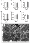
Figure 1
Figure 1
Basal characterization of Dtnbp1_KO mice. Morphometric characterization comparing parameters of 3-month-old Dtnbp1_KO and wild-type mice (n = 6 each) for heart wt.: body wt. (A), lung wt.: body wt. (B), percentage of left ventricle ejection fraction (C), percentage of left ventricle fractional shortening (D). Expression of hypertrophic genes Nppa (E) and Nppb (F) was determined by quantitative real-time PCR. (G) Electron microscopic images at various optical magnifications showing the architecture of cardiac muscle. ID: intercalated disc; Mi: mitochondria. Statistical significance was calculated by Student’s t-test. Error bars show mean ± S.E.
Figure 2
Figure 2
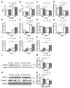
Characterization of Dtnbp1_KO mice in biomechanical stress-induced cardiomyopathy. TAC or sham operations were performed on 8-week old wild-type (WT) and Dtnbp1_KO mice. Post two weeks of operations (n = 7 (WT-SHAM), 8 (WT-TAC), 8 (KO-SHAM), 8 (KO-TAC)). Morphometric characterization showing ratios of heart weight (wt):body wt (A) and lung wt:body wt (B), functional characterization using the percentage of left ventricle ejection fraction (C) and percentage of left ventricle fractional shortening (D). Expression of hypertrophic genes Nppa (E), Nppb (F), myosin heavy chain (Myh7) (G), myosin light chain (Myh6) (H) and fibrotic markers Col1a (I), Col3a (J) and Col4a (K) was determined by quantitative real-time PCR. Representative Immunoblots display cardiac levels of SERC2A (L), with its densitometry analysis in (M). Representative Immunoblots display cardiac levels of ERK1/2 and pERK1/2 (N), with its densitometry analysis in (O). Statistical significance was calculated by two-way ANOVA. Error bars show mean ± S.E. ns, non-significant; *, p < 0.05; **, p < 0.01; ***, p < 0.001.
Figure 3
Figure 3
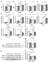
Characterization of Dtnbp1_KO mice in pharmacologically induced alpha-adrenergic stimulation -induced cardiomyopathy. 8-week old wild-type (WT) and Dtnbp1_KO mice underwent PE or PBS (control) introduction using osmotic minipumps implantation. Post two weeks of implant (n = 4 (WT-PBS), 5 (WT- PE), 5 (KO- PBS), 10 (KO- PE)), phenotypic characterization was performed by measuring morphometric characters, ratios of heart weight (wt): body wt (A), lung wt: body wt (B), functional characters like percentage of left ventricle ejection fraction (C) and percentage of left ventricle fractional shortening (D). Expression of hypertrophic genes Nppa (E), Nppb (F), myosin heavy chain (G), myosin light chain (H) and fibrotic markers Col1a (I), Col3a (J) and Col4a (K) was determined by quantitative real-time PCR. Representative Immunoblots display cellular levels of SERC2A (L), with its densitometry analysis in (M). Representative Immunoblots display cardiac levels of ERK1/2 and pERK1/2 (N), with its densitometry analysis in (O). Statistical significance was calculated by two-way ANOVA. Error bars show mean ± S.E. *, p < 0.05; **, p < 0.01; ***, p < 0.001.
Figure 4
Figure 4
Myozap and BLOC-1 complex are dysregulated after knock-out of Dysbindin. (A) Immunoblots depicting Myozap protein levels in Sham/TAC operated mice, its densitometric analysis against Tubulin shown in (B) and transcript levels in (C). (D) Immunoblots depicting the protein levels of BLOC-1 components Muted and Pallidin along with their densitometric analysis against Tubulin shown in (E) and (F), respectively. Statistical significance was calculated by two-way ANOVA. Error bars show mean ± S.E. *, p < 0.05; **, p < 0.01; ***, p < 0.001.

Similar articles

Cited by

References

    1. Nakamura M., Sadoshima J. Mechanisms of physiological and pathological cardiac hypertrophy. Nat. Rev. Cardiol. 2018;15:387–407. doi: 10.1038/s41569-018-0007-y. - DOI - PubMed
    1. Bernardo B.C., Weeks K.L., Pretorius L., McMullen J.R. Molecular distinction between physiological and pathological cardiac hypertrophy: Experimental findings and therapeutic strategies. Pharmacol. Ther. 2010;128:191–227. doi: 10.1016/j.pharmthera.2010.04.005. - DOI - PubMed
    1. Shimizu I., Minamino T. Physiological and pathological cardiac hypertrophy. J. Mol. Cell. Cardiol. 2016;97:245–262. doi: 10.1016/j.yjmcc.2016.06.001. - DOI - PubMed
    1. Yalcin F., Kucukler N., Cingolani O., Mbiyangadu B., Sorensen L., Pinherio A., Abraham M.R., Abraham T.P. Evolution of ventricular hypertrophy and myocardial mechanics in physiological and pathological hypertrophy. J. Appl. Physiol. 2019;126:354–362. doi: 10.1152/japplphysiol.00199.2016. - DOI - PMC - PubMed
    1. Niu Z., Li A., Zhang S.X., Schwartz R.J. Serum response factor micromanaging cardiogenesis. Curr. Opin. Cell Biol. 2007;19:618–627. doi: 10.1016/j.ceb.2007.09.013. - DOI - PMC - PubMed

Publication types

MeSH terms

LinkOut - more resources