. 2020 Oct 31;21(21):8158.
doi: 10.3390/ijms21218158.

Assessment of Polyethylene Glycol-Coated Gold Nanoparticle Toxicity and Inflammation In Vivo Using NF-κB Reporter Mice

Affiliations

Assessment of Polyethylene Glycol-Coated Gold Nanoparticle Toxicity and Inflammation In Vivo Using NF-κB Reporter Mice

Tzu-Yin Chen et al. Int J Mol Sci. .

Abstract

Polyethylene glycol (PEG) coating of gold nanoparticles (AuNPs) improves AuNP distribution via blood circulation. The use of PEG-coated AuNPs was shown to result in acute injuries to the liver, kidney, and spleen, but long-term toxicity has not been well studied. In this study, we investigated reporter induction for up to 90 days in NF-κB transgenic reporter mice following intravenous injection of PEG-coated AuNPs. The results of different doses (1 and 4 μg AuNPs per gram of body weight), particle sizes (13 nm and 30 nm), and PEG surfaces (methoxyl- or carboxymethyl-PEG 5 kDa) were compared. The data showed up to 7-fold NF-κB reporter induction in mouse liver from 3 h to 7 d post PEG-AuNP injection compared to saline-injected control mice, and gradual reduction to a level similar to control by 90 days. Agglomerates of PEG-AuNPs were detected in liver Kupffer cells, but neither gross pathological abnormality in liver sections nor increased activity of liver enzymes were found at 90 days. Injection of PEG-AuNPs led to an increase in collagen in liver sections and elevated total serum cholesterol, although still within the normal range, suggesting that inflammation resulted in mild fibrosis and affected hepatic function. Administrating PEG-AuNPs inevitably results in nanoparticles entrapped in the liver; thus, further investigation is required to fully assess the long-term impacts by PEG-AuNPs on liver health.

Keywords: NF-κB; PEG surface modification; gold nanoparticle; liver inflammation; reporter imaging; steatosis.


Conflict of interest statement

The authors declare no conflict of interest. The funders had no role in the design of the study; in the collection, analyses, or interpretation of data; in the writing of the manuscript, or in the decision to publish the results.

Figures

Figure 1
Figure 1
NF-κB dual reporter mouse and in vivo response to LPS and TNF-α treatment. (a) Schematic of the transgenic vector (not to scale). An NF-κB responsive promoter was placed before the coding sequence of firefly luciferase (fLuc) and the herpes simplex virus truncated thymidine kinase (HSV1Δtk) fusion protein. (b) Bioluminescence imaging of reporter mice before (0 h), and at 4 h, 8 h, and 24 h after i.p. injection of LPS (1 mg/Kg) or TNF-α (2 μg). (c) Fold induction of bioluminescence by LPS or TNF-α within the abdominal region of interest (ROI) compared to pre-injection values; n = 3. LPS, lipopolysaccharide.
Figure 2
Figure 2
Hydrodynamic and core sizes of methoxyl- or carboxymethyl-PEG5K coated AuNPs. (a,c) The hydrodynamic diameter, Zave, of 13 nm mPEG-AuNPs and 30 nm cPEG-AuNPs, respectively, determined by dynamic light scattering spectroscopy. (b,d) TEM imaging of 13 nm mPEG-AuNPs and 30 nm cPEG-AuNPs. (e) Sizes and ζ potentials of 13 nm and 30 nm PEG-AuNPs, averaged from five and four batch preparations, respectively. AuNP, gold nanoparticle; PEG, polyethylene glycol; TEM, transmission electron microscopy.
Figure 3
Figure 3
Bioluminescence in reporter mice in response to intravenous PEGylated AuNP injection. (a) Low (1 μg/g) or high (4 μg/g) doses of 13 or 30 nm mPEG- (M13, M30) or cPEG-AuNPs (C13, C30) were injected into mice. Images were acquired at the indicated time points for up to 3 months (mos) under the same conditions and imaging settings. (b) Mean radiance (photons/sec/cm2/sr) of the liver ROI of mice injected with high dose PEG-AuNPs; n = 3; * p < 0.05 compared to groups at 3 mos post-injection with C13, M30, C30, or saline.
Figure 4
Figure 4
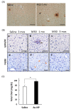
Detection of p65 nuclear localization, Ly6G/6C positive monocytes, and F4/80 positive macrophages in liver, and increased total serum cholesterol. (A) p65 immunostaining of liver sections with an anti-p65 antibody and nuclear counterstaining with methyl green. Mouse liver sections from 1 mo post-injection with 13 nm mPEG-AuNPs or with saline are shown. Clusters of dark brown color include positively stained cells and some dye debris. Nuclear-localized p65 was found in Kupffer cells (white arrow) and hepatocytes (black arrow). Scale bar = 50 μm. (B) Ly6G/6C positive monocytes (ac) and F4/80 positive macrophages (df) were readily detected in liver sections at 1 mo post-injection with 30 nm mPEG-AuNPs (black arrow in b; orange arrows in e), but rarely detected in liver samples harvested at 3 mos post-injection (c, f) or in those from saline-injected control mice (a,d). Scale bar = 50 μm. (C) Total serum cholesterol (Total Chol) in Tg mice injected with high doses of cPEG- or mPEG-AuNPs (n = 19) was significantly higher than that in the saline-injected group (n = 8); * p < 0.05.
Figure 5
Figure 5
Agglomerates of PEG-AuNPs in liver Kupffer cells at 3 mos post 13 nm mPEG- or cPEG-AuNP injection (a,b), or by 30 nm mPEG- or cPEG-AuNP injection (c,d). Higher magnification images are shown at right.
Figure 6
Figure 6
Pathology and collagen deposition in liver tissue from a PEG-AuNP-injected mouse. (A) H&E (af) and Sirius Red (gi) staining of liver sections of control (a,d,g), and at 1 mo (b,e,h), and 3 mos (c,f,i) after injection with 30 nm mPEG-AuNPs (4 μg/g). There were no significant abnormalities due to nanoparticle injection in liver sections (af). Siris Red staining of sinusoidal microvasculature increased at 1 mo and 3 mos after PEG-AuNP injection compared to saline-injected controls (gi). Scale bar = 100 μm. (B) Semiquantitative estimation of collagen content in liver sections via the ratio of dye absorbances of Sirius Red (540 nm) and Fast Green (605 nm) after dye extraction from stained sections. C: saline injection.

Similar articles

Cited by

References

    1. Cho W.-S., Cho M., Jeong J., Choi M., Cho H.-Y., Han B.S., Kim S.H., Kim H.O., Lim Y.T., Chung B.H., et al. Acute toxicity and pharmacokinetics of 13 nm-sized PEG-coated gold nanoparticles. Toxicol. Appl. Pharmacol. 2009;236:16–24. doi: 10.1016/j.taap.2008.12.023. - DOI - PubMed
    1. Zhang J., Nie X., Ji Y., Liu Y., Wu X., Chen C., Fang X. Quantitative biokinetics and systemic translocation of various gold nanostructures are highly dependent on their size and shape. J. Nanosci. Nanotechnol. 2014;14:4124–4138. doi: 10.1166/jnn.2014.8274. - DOI - PubMed
    1. De Jong W.H., Hagens W.I., Krystek P., Burger M.C., Sips A.J., Geertsma R.E. Particle size-dependent organ distribution of gold nanoparticles after intravenous administration. Biomaterials. 2008;29:1912–1919. doi: 10.1016/j.biomaterials.2007.12.037. - DOI - PubMed
    1. Li B., Lane L.A. Probing the biological obstacles of nanomedicine with gold nanoparticles. WIREs Nanomed. Nanobiotechnol. 2019;11:e1542. doi: 10.1002/wnan.1542. - DOI - PMC - PubMed
    1. Lee K.Y., Lee G.Y., Lane L.A., Li B., Wang J., Lu Q., Wang Y., Nie S. Functionalized, long-circulating, and ultrasmall gold nanocarriers for overcoming the barriers of low nanoparticle delivery efficiency and poor tumor penetration. Bioconjug. Chem. 2017;28:244–252. doi: 10.1021/acs.bioconjchem.6b00224. - DOI - PubMed

LinkOut - more resources