. 2020 Oct 31;8(11):1707.
doi: 10.3390/microorganisms8111707.

Origin and Evolution of Studiervirinae Bacteriophages Infecting Pectobacterium: Horizontal Transfer Assists Adaptation to New Niches

Affiliations

Origin and Evolution of Studiervirinae Bacteriophages Infecting Pectobacterium: Horizontal Transfer Assists Adaptation to New Niches

Peter V Evseev et al. Microorganisms. .

Abstract

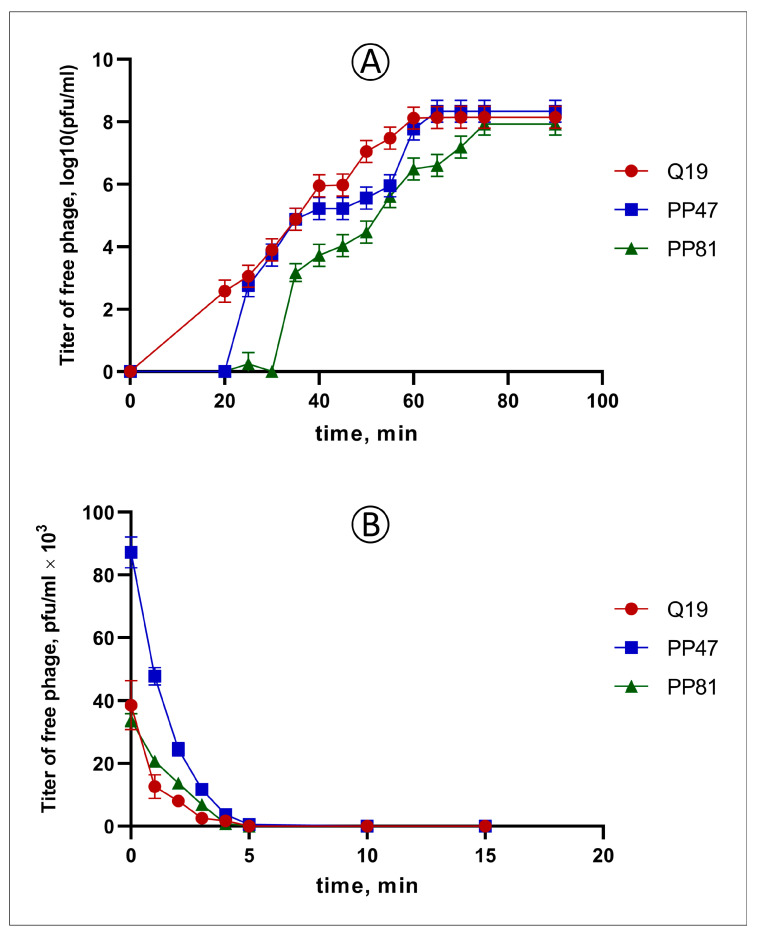
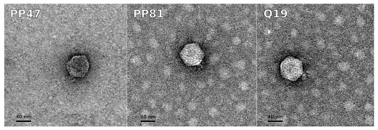
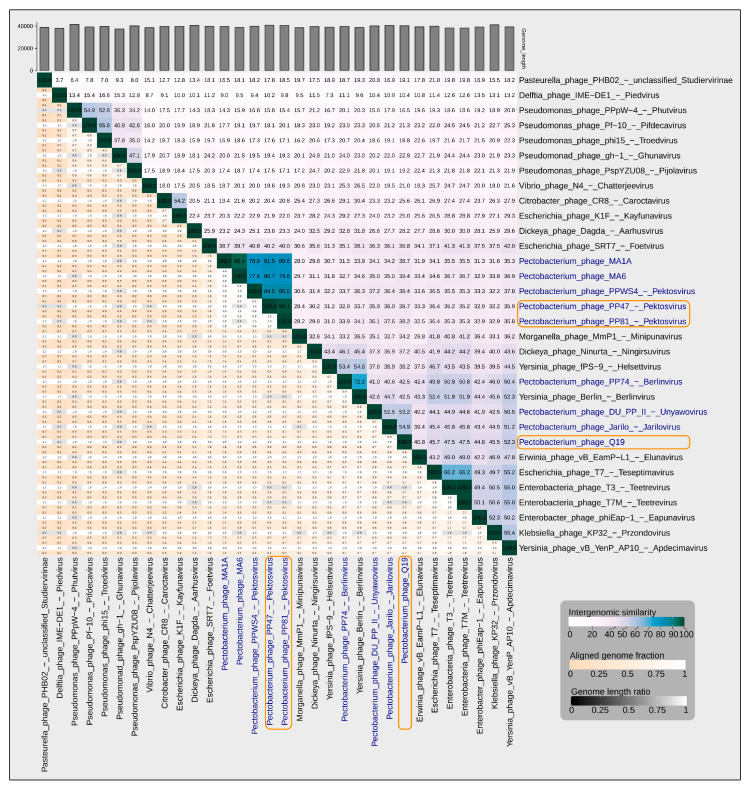
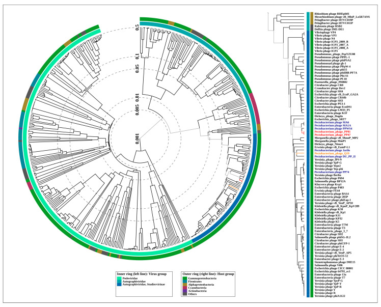
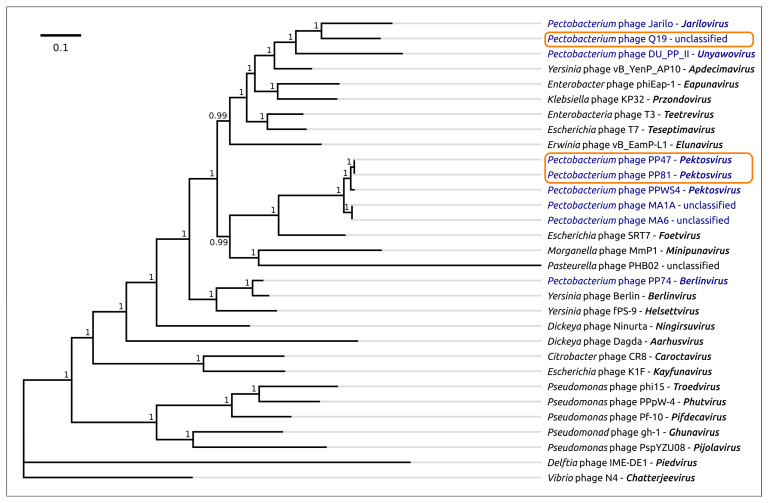
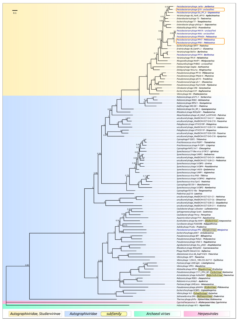
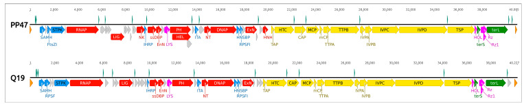
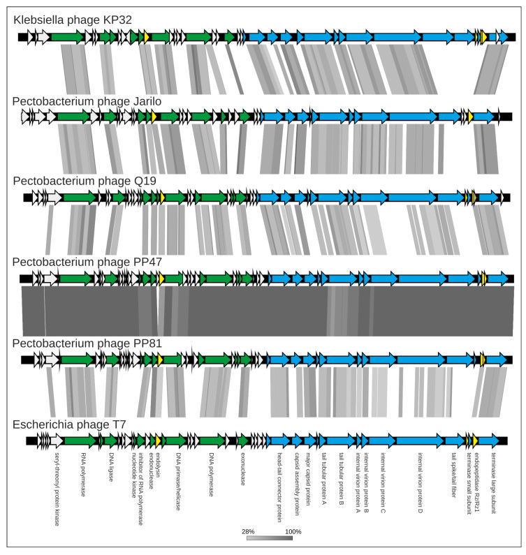
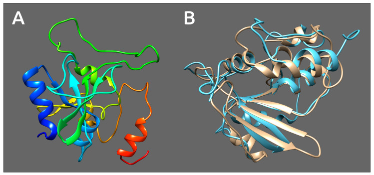
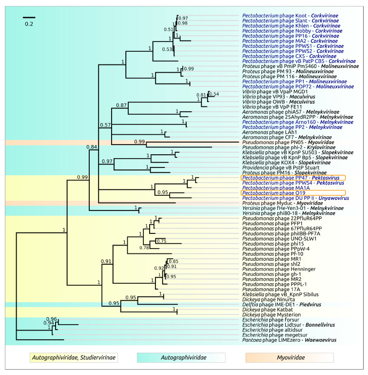
Black leg and soft rot are devastating diseases causing up to 50% loss of potential potato yield. The search for, and characterization of, bacterial viruses (bacteriophages) suitable for the control of these diseases is currently a sought-after task for agricultural microbiology. Isolated lytic Pectobacterium bacteriophages Q19, PP47 and PP81 possess a similar broad host range but differ in their genomic properties. The genomic features of characterized phages have been described and compared to other Studiervirinae bacteriophages. Thorough phylogenetic analysis has clarified the taxonomy of the phages and their positioning relative to other genera of the Autographiviridae family. Pectobacterium phage Q19 seems to represent a new genus not described previously. The genomes of the phages are generally similar to the genome of phage T7 of the Teseptimavirus genus but possess a number of specific features. Examination of the structure of the genes and proteins of the phages, including the tail spike protein, underlines the important role of horizontal gene exchange in the evolution of these phages, assisting their adaptation to Pectobacterium hosts. The results provide the basis for the development of bacteriophage-based biocontrol of potato soft rot as an alternative to the use of antibiotics.

Keywords: Autographiviridae; Pectobacterium; evolution; horizontal transfer; phage; phage therapy; tail spike.


Conflict of interest statement

The authors declare no conflict of interest. The funders had no role in the design of the study; in the collection, analyses, or interpretation of data; in the writing of the manuscript; or in the decision to publish the results.

Figures

Figure 1
Figure 1
(A) One-step growth curve of phages Q19 (red), PP47 (blue) and PP81 (green) using P. brasiliense F157 as host strain in MOI = 0.01. (B) Adsorption of phages Q19 (red), PP47 (blue) and PP81 (green) at the surface of P. brasiliense F157 in MOI = 0.001.
Figure 2
Figure 2
Transmission electron microscopy of bacteriophages PP47, PP81, and Q19. Staining was with 1% uranyl acetate. The scale bar is 40 nm.
Figure 3
Figure 3
VIRIDIC generated heatmap of 30 Studiervirinae phages and a representative of the Piedvirus genus, which is closely related to Studiervirinae. The heatmap incorporates intergenomic similarity values (right half) and alignment indicators (left half and top annotation). In the right half, the colour coding allows a rapid visualisation of the clustering of the phage genomes based on intergenomic similarity. The numbers represent the similarity values for each genome pair, rounded to the first decimal. In the left half, three indicator values are represented for each genome pair, from top to bottom: aligned fraction genome 1 (for the genome found in this row), genome length ratio (for the two genomes in this pair) and aligned fraction genome 2 (for the genome found in this column). Pectobacterium phages PP47, PP81, PPWS4, MA6 and MA1A are clustered with an intergenomic similarity higher than the genus threshold of 70%. Pectobacterium phage Q19 has an intergenomic similarity that is lower than 70% compared to any other phage.
Figure 4
Figure 4
Circular proteomic tree of 447 Podovoridae and Autographiviridae phage genomes, and the Studiervirinae part of the tree (right) constructed using ViPTree. The branches representing Pectobacterium phages PP47 and PP81 are coloured red, and the branch representing Pectobacterium phage Q19 is coloured orange.
Figure 5
Figure 5
Phylogenetic tree obtained with MrBayes, based on concatenated sequences of DNA polymerase, a large subunit of terminase, a head-tail connector protein, a major capsid protein and a single-strand DNA binding protein extracted from the genomes of 29 phages, recognised by International Committee on Taxonomy of Viruses (ICTV) as master species belonging to the Studiervirinae subfamily, Pectobacterium phage PP74 of the Berlinvirus genus and Delphia phage IME-DE1. Bayesian posterior probabilities are indicated above their branch. Taxonomic classification is taken from ICTV and is shown to the right of the organism name. The scale bar shows 0.1 estimated substitutions per site and the tree was rooted to Delphia phage IME-DE1; of 2,000,000 generations, every 200 generations were sampled, with an average standard deviation of split frequencies of 0.0027.
Figure 6
Figure 6
Phylogenetic tree obtained with MrBayes based on 100 terminase large subunit protein sequences. Bayesian posterior probabilities are indicated above their branch. Taxonomic classification is taken from ICTV and is shown to the right of the organism name. The scale bar shows 0.1 estimated substitutions per site and the tree was rooted to Cyprinid herpesvirus 3; of 2,000,000 generations, every 200 generations were sampled, with an average standard deviation of split frequencies of 0.012.
Figure 7
Figure 7
Genetic map of phages PP47 and Q19. The colors of different functional modules are as follows: yellow, morphogenesis; red, replication, transcription and nucleic acids processing; green, packaging; purple, lysis proteins; blue, regulation of host defense and metabolic processes; orange, terminal repeats; grey, hypothetical proteins. Putative transcriptional promoters, predicted with Phage Promoters, are shown above each sequence (presented as a black line) and colored cyan. Numbers above the sequences show the position in genomes. Genes’ names are as follows: SAMH, S-adenosyl-L-methionine hydrolase; RPSF, RNA polymerase σ54 factor; FtsZI, cell division FtsZ inhibitor; STPK, seryl-threonyl protein kinase; RNAP, DNA-directed RNA polymerase; LIG, DNA ligase; NK, nucleotide kinase; IHRP, inhibitor of host bacterial RNA polymerase; ssDBP, ssDNA-binding protein CDS; EnN, endonuclease I; LYS, lysozyme, N-acetylmuramoyl-L-alanine amidase; PH, DNA primase/helicase; HEL, DNA helicase; ITA, inhibitor of toxin/antitoxin system; NT, nucleotidyltransferase; DNAP, DNA polymerase; HNSBP, H-NS and tRNA binding protein CDS; RPSFI, host RNA polymerase σ70 factor inhibitor; ExN, 5′-3′ exonuclease; HNH, HNH endonuclease; TAP, tail assembly protein; HTC, head-tail connector protein; CAP, capsid assembly protein; MCP, major capsid protein; mCP, minor capsid protein; TTPA, tail tubular protein A; TTPB, tail tubular protein B; IVPA, internal virion protein A; IVPB, internal virion protein B; IVPC, internal virion protein C; IVPD, internal virion protein D; TSP, tail spike protein, SGNH hydrolase domain-containing protein; HOL, class II holin; terS, terminase small subunit; Rz, Rz lysis protein; Rz1, Rz1 lysis protein; terL, terminase large subunit.
Figure 8
Figure 8
Genome sequence comparison among six Studiervirinae viral genomes exhibiting co-linearity detected by TBLASTX. The percentage of sequence similarity is indicated by the intensity of the grey color. Vertical blocks between analyzed sequences indicate regions with at least 28% similarity. Nucleic acid-processing genes are colored green, morphogenesis and packaging genes are colored blue and lysis genes are colored yellow. The most significant differences are observed for tail proteins and a number of hypothetical proteins of early and middle regions.
Figure 9
Figure 9
Upper picture: 3D homology modelling of tail spike proteins of Pectobacterium phages Q19, PP47 and PP81, and PDB structure of SGNH domain of bacterial esterase (CEX) active on acetylated mannans PDB structure 6hfz. Lower picture: 3D-alignment of modelled Q19, PP47 and PP81 tail spike SGNH domains, with SGNH domain of bacterial esterase (CEX) as a template. In the upper picture, the five parallel β-sheets intrinsic for all SGNH are colored yellow; the other β-sheets are colored blue, α-helices are colored red and coils are colored green.
Figure 10
Figure 10
Phylogenetic tree obtained with MrBayes, based on amino acid sequences of the N-domain of tail fibre/tail spike proteins and homologous sequences obtained by a BLAST search of Genbank phage and bacterial databases. Bayesian posterior probabilities are indicated above their branch. The scale bar shows 0.05 estimated substitutions per site and the tree was rooted to Megasphaera hexanoica; of 2,000,000 generations, 200 generations were sampled, with an average standard deviation of split frequencies of 0.011.
Figure 11
Figure 11
Phylogenetic tree obtained with MrBayes, based on amino acid sequences of the central domain of tail fibre/tail spike proteins and homologous sequences obtained by a BLAST search of Genbank phage and bacterial databases. Bayesian posterior probabilities are indicated above their branch. The scale bar shows 0.2 estimated substitutions per site and the tree was rooted to Sinorhizobium meliloti BL225C plasmid; of 2,000,000 generations, 200 generations were sampled, with an average standard deviation of split frequencies of 0.0062.
Figure 12
Figure 12
(A) Simplified genetic maps of Pectobacterium Studiervirinae phages PP47, PP81, PPWS4, MA6, MA1A, Q19, Jarilo, DU_PP_II and PP74, and phages PP1 and POP72. (B) Regions of Pectobacterium aroidearum PC1 and Pectobacterium polaris PZ1 containing the genes of SGNH-domain proteins, homologous to Q19 and PP47/81 tail spike proteins. The colors of different genes are as follows: orange, tail spike protein; green, tail fibre protein; cyan, tail tubular protein B; blue, holin; dark-blue, HNH endonuclease; yellow, nucleotidyltransferase; purple, recombination protein RecR and conjugal transfer protein TraB; grey, other genes. Putative functions of genes are given according to Genbank annotations and BLAST search (Tables S6–S8).
Figure 13
Figure 13
(A) 3D structure homology modelling of tRNA-nycleotidyltransferase from Pectobacterium phage PP47. The model is colored based on a rainbow gradient scheme, where the N-terminus of the polypeptide chain is colored blue and the C-terminus is colored red. (B) 3D-alignment of modelled tRNA-nycleotidyltransferase from Pectobacterium phage PP47 (colored blue) and Pectobacerium aroidearum PC1 (colored sand).
Figure 14
Figure 14
Phylogenetic tree obtained with MrBayes, based on amino acid sequences of tRNA-nucleotidyltransferase and homologous sequences obtained by a BLAST search of Genbank phage databases. Bayesian posterior probabilities are indicated above their branch. The scale bar shows 0.2 estimated substitutions per site and the tree was rooted to Pantoea phage LIMEzero; of 2,000,000 generations, every 200 generations were sampled, with an average standard deviation of split frequencies of 0.0071.

Similar articles

Cited by

References

    1. Czajkowski R., Pérombelon M.C.M., Van Veen J.A., Van der Wolf J.M. Control of blackleg and tuber soft rot of potato caused by Pectobacterium and Dickeya species: A review. Plant. Pathol. 2011;60:999–1013. doi: 10.1111/j.1365-3059.2011.02470.x. - DOI
    1. Pérombelon M.C.M. Potato diseases caused by soft rot erwinias: An overview of pathogenesis. Plant. Pathol. 2002;51:1–12.
    1. Ma B., Hibbing M.E., Kim H.-S., Reedy R.M., Yedidia I., Breuer J.J., Breuer J.J., Glasner J.D., Perna N.T., Kelman A., et al. Host range and molecular phylogenies of the soft rot enterobacterial genera pectobacterium and dickeya. Phytopathology. 2007;97:1150–1163. doi: 10.1094/PHYTO-97-9-1150. - DOI - PubMed
    1. Motyka A., Zoledowska S., Sledz W., Lojkowska E. Molecular methods as tools to control plant diseases caused by Dickeya and Pectobacterium spp: A minireview. New Biotechnol. 2017;39:181–189. doi: 10.1016/j.nbt.2017.08.010. - DOI - PubMed
    1. Svircev A., Roach D., Castle A. Framing the future with bacteriophages in agriculture. Viruses. 2018:218. doi: 10.3390/v10050218. - DOI - PMC - PubMed

LinkOut - more resources