. 2020 Oct 31;21(21):8171.
doi: 10.3390/ijms21218171.

Single-Cell Analysis of Different Stages of Oral Cancer Carcinogenesis in a Mouse Model

Affiliations

Single-Cell Analysis of Different Stages of Oral Cancer Carcinogenesis in a Mouse Model

Ling-Yu Huang et al. Int J Mol Sci. .

Abstract

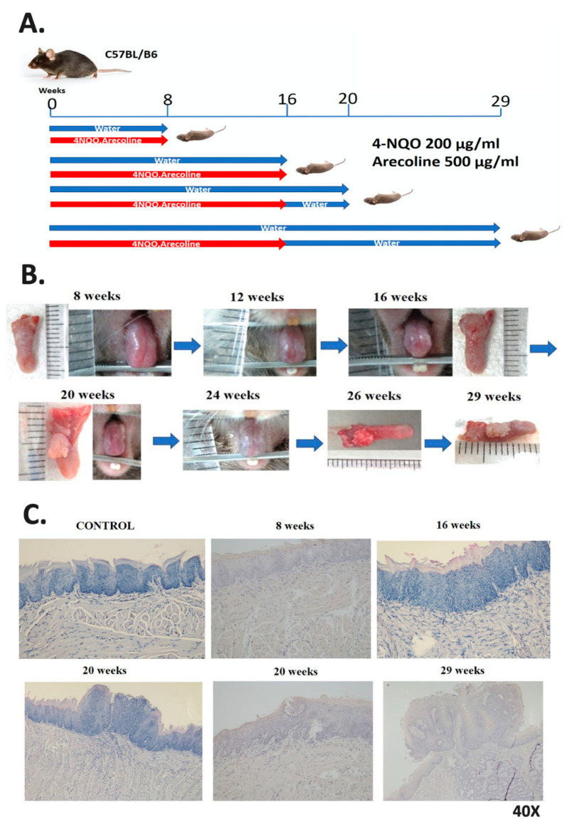
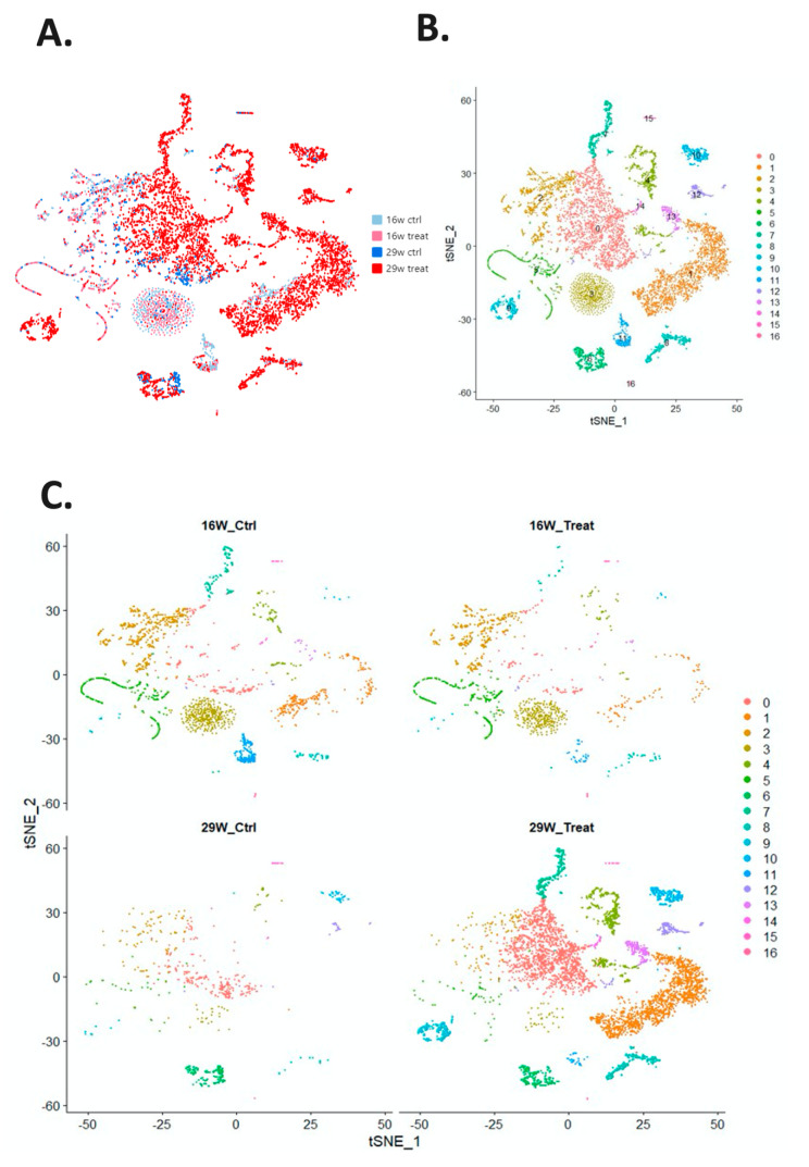
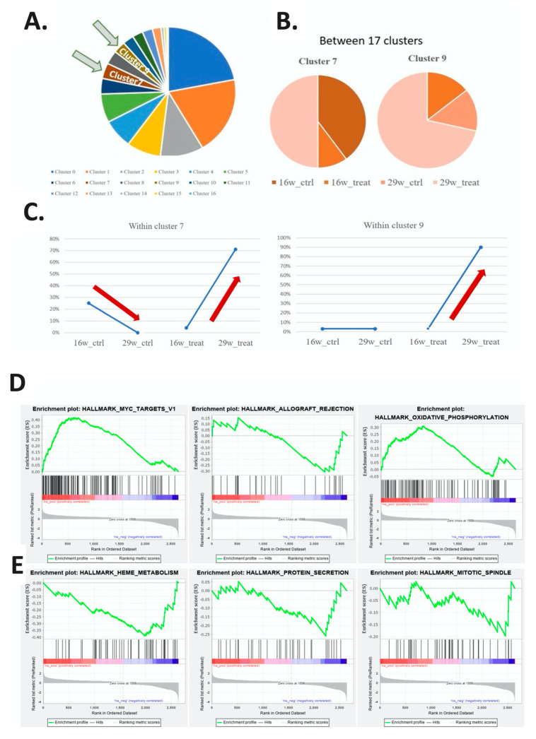
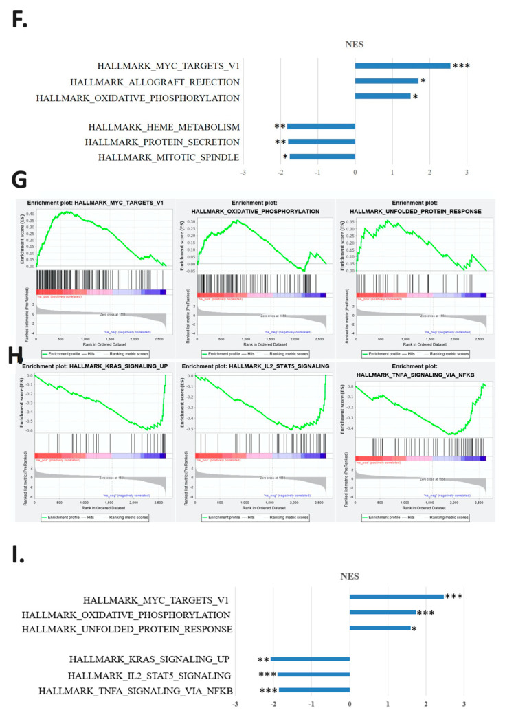
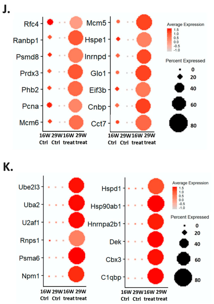
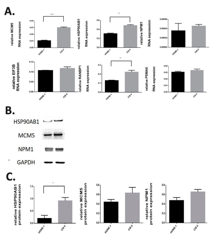
Oral carcinogenesis involves the progression of the normal mucosa into potentially malignant disorders and finally into cancer. Tumors are heterogeneous, with different clusters of cells expressing different genes and exhibiting different behaviors. 4-nitroquinoline 1-oxide (4-NQO) and arecoline were used to induce oral cancer in mice, and the main factors for gene expression influencing carcinogenesis were identified through single-cell RNA sequencing analysis. Male C57BL/6J mice were divided into two groups: a control group (receiving normal drinking water) and treatment group (receiving drinking water containing 4-NQO (200 mg/L) and arecoline (500 mg/L)) to induce the malignant development of oral cancer. Mice were sacrificed at 8, 16, 20, and 29 weeks. Except for mice sacrificed at 8 weeks, all mice were treated for 16 weeks and then either sacrificed or given normal drinking water for the remaining weeks. Tongue lesions were excised, and all cells obtained from mice in the 29- and 16-week treatment groups were clustered into 17 groups by using the Louvain algorithm. Cells in subtypes 7 (stem cells) and 9 (keratinocytes) were analyzed through gene set enrichment analysis. Results indicated that their genes were associated with the MYC_targets_v1 pathway, and this finding was confirmed by the presence of cisplatin-resistant nasopharyngeal carcinoma cell lines. These cell subtype biomarkers can be applied for the detection of patients with precancerous lesions, the identification of high-risk populations, and as a treatment target.

Keywords: carcinogenesis; mouse model; oral cancer; single-cell RNA sequencing.


Conflict of interest statement

The authors declare no conflict of interest.

Figures

Figure 1
Figure 1
The carcinogen 4-nitroquinoline 1-oxide (4-NQO) (similar to carcinogens found in cigarettes) and arecoline (the carcinogen found in betel nut) were used to induce head and neck cell carcinogenesis in mice. (A) Four experimental and control groups were examined. Mice in the experimental group were fed drinking water containing 4-NQO and arecoline and then monitored until being sacrificed. (B) The progression of precancerous lesions on the tongue to cancer was monitored when mice were alive, and they were sacrificed at different time points. With the increasing duration of treatment in the experimental group, white lesions began to appear on the tongues of mice. Tumors were observed on the tongues of mice at week 16 and had become invasive carcinoma by week 29. (C) The tongues of mice in the control and experimental groups underwent hematoxylin and eosin staining for histopathological examination.
Figure 2
Figure 2
Visual distribution of dimensionality reduction in cell populations. (A) The cell distribution of all samples generated through t-distributed stochastic neighbor embedding and t-random neighbor embedding, with four groups as the source (16- and 29-week control and experimental groups). (B) Cells divided into 17 subtypes by using the Louvain algorithm. (C) Separation of the four major groups of cell populations and the distribution of the 17 cell subtypes. (D) Heat map of the 17 cell subtypes. After cells were separated into 17 subtypes, the 10 genes with the highest expression in each subtype of cells were identified and compared between subtypes. Yellow indicates that the gene expression is higher than in other cell subtypes.
Figure 2
Figure 2
Visual distribution of dimensionality reduction in cell populations. (A) The cell distribution of all samples generated through t-distributed stochastic neighbor embedding and t-random neighbor embedding, with four groups as the source (16- and 29-week control and experimental groups). (B) Cells divided into 17 subtypes by using the Louvain algorithm. (C) Separation of the four major groups of cell populations and the distribution of the 17 cell subtypes. (D) Heat map of the 17 cell subtypes. After cells were separated into 17 subtypes, the 10 genes with the highest expression in each subtype of cells were identified and compared between subtypes. Yellow indicates that the gene expression is higher than in other cell subtypes.
Figure 3
Figure 3
Comparison of the compositions of cell subtypes between the 16- and 29-week control and experimental groups. (A) Proportion of the 17 cell subtypes with respect to the total cell composition presented as a pie chart. (B) Comparison of the proportion of the seventh and ninth cell subtypes in the four major groups. (C) Line graph comparing the proportions of the seventh and ninth cell subtypes in the four major groups. The proportion increased with increasing treatment duration in in the experimental group. (DF) Gene function enrichment analysis of the seventh cell subtype. (D) Line chart indicating the three most significant pathways involved in the increase in gene expression in the 16- and 29-week experimental groups. (E) Line chart showing the three most significant pathways involved in the decline in gene expression in the 16- and 29-week experimental groups. (F) Bar graph of the normalized enrichment scores (NESs) and p values of these three paths (* p < 0.05; ** p < 0.01; *** p < 0.001). (GI) Gene function enrichment analysis of the ninth cell subtype. (G) Line chart indicating the three most significant pathways involved in the increase in gene expression in the 16- and 29-week experimental groups. (H) Line chart showing three most significant pathways involved in the decline in gene expression in the 16- and 29-week experimental groups. (I) Bar graph displaying the calculated NESs and p values of these three paths (* p < 0.05; ** p < 0.01; *** p < 0.001). (J) Dot diagram of genes involved in the MYC_targets_v1 pathway in the seventh subtype. The average gene expression level and the percentage of cells in the four groups are indicated by the color and size of dots. The average expression level and cell ratio of genes in the 29-week experimental group were significantly higher than those in the other groups. (K) Dot diagram of genes involved in the MYC_targets_v1 pathway in the ninth subtype. The average gene expression level and the percentage of cells in the four groups are indicated by the color and size of dots. The average expression level and cell ratio of genes in the 29-week experimental group were significantly higher than those in the other groups.
Figure 3
Figure 3
Comparison of the compositions of cell subtypes between the 16- and 29-week control and experimental groups. (A) Proportion of the 17 cell subtypes with respect to the total cell composition presented as a pie chart. (B) Comparison of the proportion of the seventh and ninth cell subtypes in the four major groups. (C) Line graph comparing the proportions of the seventh and ninth cell subtypes in the four major groups. The proportion increased with increasing treatment duration in in the experimental group. (DF) Gene function enrichment analysis of the seventh cell subtype. (D) Line chart indicating the three most significant pathways involved in the increase in gene expression in the 16- and 29-week experimental groups. (E) Line chart showing the three most significant pathways involved in the decline in gene expression in the 16- and 29-week experimental groups. (F) Bar graph of the normalized enrichment scores (NESs) and p values of these three paths (* p < 0.05; ** p < 0.01; *** p < 0.001). (GI) Gene function enrichment analysis of the ninth cell subtype. (G) Line chart indicating the three most significant pathways involved in the increase in gene expression in the 16- and 29-week experimental groups. (H) Line chart showing three most significant pathways involved in the decline in gene expression in the 16- and 29-week experimental groups. (I) Bar graph displaying the calculated NESs and p values of these three paths (* p < 0.05; ** p < 0.01; *** p < 0.001). (J) Dot diagram of genes involved in the MYC_targets_v1 pathway in the seventh subtype. The average gene expression level and the percentage of cells in the four groups are indicated by the color and size of dots. The average expression level and cell ratio of genes in the 29-week experimental group were significantly higher than those in the other groups. (K) Dot diagram of genes involved in the MYC_targets_v1 pathway in the ninth subtype. The average gene expression level and the percentage of cells in the four groups are indicated by the color and size of dots. The average expression level and cell ratio of genes in the 29-week experimental group were significantly higher than those in the other groups.
Figure 4
Figure 4
RNA and protein expression in cisplatin-resistant nasopharyngeal carcinoma cell lines. (A) Expression of MCM5, HSP90AB1, NPM1, EIF3B, RANBP1, and PSMA6 RNA analyzed through real-time quantitative polymerase chain reaction in the nasopharyngeal carcinoma cell line HONE-1 and the cisplatin-resistant nasopharyngeal carcinoma cell line HONE1-CIS6. Expression levels were higher in the HONE1-CIS6 cell line than in the nasopharyngeal carcinoma cell lines (** p < 0.005; **** p < 0.0001). (B) Protein expression levels of HSP90AB1, MCM5 and NPM1 analyzed in HONE-1 and HONE1-CIS6 through Western blotting; HONE1-CIS6 had the highest protein expression levels (* p < 0.05). (C) Relative quantitative protein expression levels of HSP90AB1, MCM5 and NPM1 to GAPDH in HONE-1 and HONE1-CIS6.

References

    1. Lousada-Fernandez F., Rapado-Gonzalez O., Lopez-Cedrun J.L., Lopez-Lopez R., Muinelo-Romay L., Suarez-Cunqueiro M.M. Liquid Biopsy in Oral Cancer. Int. J. Mol. Sci. 2018;19:1704. doi: 10.3390/ijms19061704. - DOI - PMC - PubMed
    1. Kumar M., Nanavati R., Modi T.G., Dobariya C. Oral cancer: Etiology and risk factors: A review. J. Cancer Res. Ther. 2016;12:458–463. doi: 10.4103/0973-1482.186696. - DOI - PubMed
    1. Wong T., Wiesenfeld D. Oral Cancer. Aust. Dent. J. 2018;63(Suppl. 1):S91–S99. doi: 10.1111/adj.12594. - DOI - PubMed
    1. Shen Y., Liu J., Zhang L., Dong S., Zhang J., Liu Y., Zhou H., Dong W. Identification of Potential Biomarkers and Survival Analysis for Head and Neck Squamous Cell Carcinoma Using Bioinformatics Strategy: A Study Based on TCGA and GEO Datasets. BioMed Res. Int. 2019;2019:7376034. doi: 10.1155/2019/7376034. - DOI - PMC - PubMed
    1. Zevallos J.P., Mazul A.L., Walter V., Hayes D.N. Gene Expression Subtype Predicts Nodal Metastasis and Survival in Human Papillomavirus-Negative Head and Neck Cancer. Laryngoscope. 2019;129:154–161. doi: 10.1002/lary.27340. - DOI - PMC - PubMed

MeSH terms