Review
. 2020 Oct 30;8(11):460.
doi: 10.3390/biomedicines8110460.

Osmotic Adaptation by Na+-Dependent Transporters and ACE2: Correlation with Hemostatic Crisis in COVID-19

Affiliations
Review

Osmotic Adaptation by Na+-Dependent Transporters and ACE2: Correlation with Hemostatic Crisis in COVID-19

Danah Muhanna et al. Biomedicines. .

Abstract

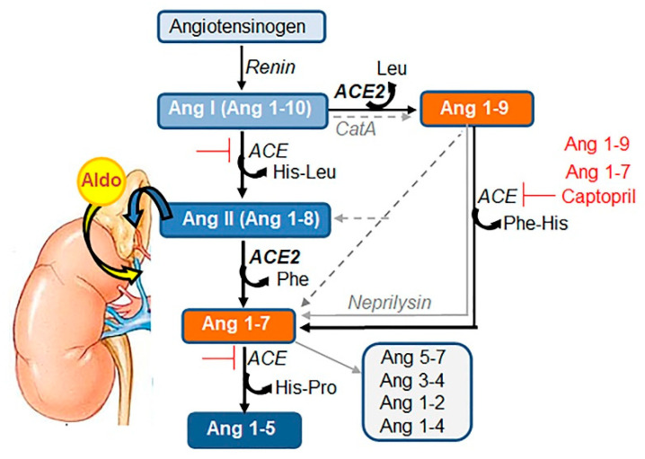
COVID-19 symptoms, including hypokalemia, hypoalbuminemia, ageusia, neurological dysfunctions, D-dimer production, and multi-organ microthrombosis reach beyond effects attributed to impaired angiotensin-converting enzyme 2 (ACE2) signaling and elevated concentrations of angiotensin II (Ang II). Although both SARS-CoV (Severe Acute Respiratory Syndrome Coronavirus) and SARS-CoV-2 utilize ACE2 for host entry, distinct COVID-19 pathogenesis coincides with the acquisition of a new sequence, which is homologous to the furin cleavage site of the human epithelial Na+ channel (ENaC). This review provides a comprehensive summary of the role of ACE2 in the assembly of Na+-dependent transporters of glucose, imino and neutral amino acids, as well as the functions of ENaC. Data support an osmotic adaptation mechanism in which osmotic and hemostatic instability induced by Ang II-activated ENaC is counterbalanced by an influx of organic osmolytes and Na+ through the ACE2 complex. We propose a paradigm for the two-site attack of SARS-CoV-2 leading to ENaC hyperactivation and inactivation of the ACE2 complex, which collapses cell osmolality and leads to rupture and/or necrotic death of swollen pulmonary, endothelial, and cardiac cells, thrombosis in infected and non-infected tissues, and aberrant sensory and neurological perception in COVID-19 patients. This dual mechanism employed by SARS-CoV-2 calls for combinatorial treatment strategies to address and prevent severe complications of COVID-19.

Keywords: angiotensin; coagulation; hypertension; inflammation; organ failure; thrombosis; tonicity; transporters; virus.


Conflict of interest statement

The authors declare no conflict of interest. The funders had no role in the design of the study; in the collection, analyses, or interpretation of data; in the writing of the manuscript, or in the decision to publish the results.

Figures

Figure 1
Figure 1
Components of the Renin-Angiotensin System (RAS). Bioactive peptides of angiotensinogen, Ang I or Ang (1–10), Ang II or Ang (1–8), and Ang (1–7) are depicted as rectangular shapes. The cleavage enzymes of renin, angiotensin-converting enzyme (ACE), ACE2, cathepsin A (CatA), and neprilysin are indicated in italics. The central panel shows the canonic RAS pathway. ACE cleaves dipeptide His-Leu, which converts decapeptide Ang I into octapeptide Ang II (blue rectangles). ACE2 cleaves a neutral amino acid, Phe, that converts Ang II into Ang 1–7 (orange rectangles). The alternative pathways by ACE and ACE2 (black arrows) as well as those mediated by other enzymes (gray lines) are shown on the right. Captopril is synthetic and Ang 1–7 and Ang 1–9 are natural inhibitors of ACE (red lines). Ang 1–7 inhibition presents a classical feedback mechanism for physiological control of Ang II levels in circulation. One example of the convergence of Ang II with other endocrine pathways for the regulation of blood pressure is shown on the left. Circulating Ang II initiates the production of aldosterone (Aldo) from cholesterol in the adrenal cortex.
Figure 2
Figure 2
Schematic regulation of ACE2-dependent pathways under physiological conditions and during SARS-CoV-2 infection. (A) Main signaling in the absence of ACE2 when it is not expressed, or its activity is inhibited (ACE2 is ‘OFF’). Circulating Ang II binds to its high affinity G-protein-coupled receptor AT1R. Main AngII/AT1R signaling mediates vasoconstriction, NADPH oxidase activation and reactive oxygen species (ROS) formation, hypoxia induction (HIF1α), activation of MMPs, including ADAM17 and pro-inflammatory cytokines, Na+/H2O retention from extracellular fluids into blood, hypertension, hypertrophy, activation of sympathetic nerves and other effects. Ang II/AT1R receptor can suppress angiotensin 2 receptor (AT2R, red lines) expression in the initial stages of inflammation, but this suppression is alleviated by interferon regulatory factor 1 (IRF-1). Ang II also activates AT2R, sometimes in complex with different proteins, which opposes the effects of Ang II/AT1R and resolves of stress, inflammation, and apoptosis. Under specific conditions, Ang II/AT2R response could mimic Ang II/AT1R possibly by involving further cleavage of Ang II. MAS receptor remains inactivated (OFF) without ligand Ang (1–7), produced from Ang II. (B) The main signaling in the presence of catalytically active ACE2 (ACE2 is ‘ON’). Activated ACE2 is shown as an integral protein with an active catalytic site containing Zn2+ in the ectodomain (green V shape). ACE2 is stabilized in a catalytically active conformation by calmodulin (CaM, red rectangle) bound to an intracellular domain of ACE2 that prevents cleavage of ACE2 by proteases ADAM17 (not shown) or TMPRSS2 (orange line shape). ACE2 cleaves Ang II, leaving both AT1R and AT2R inactive (OFF). ACE2 cleavage produces Ang 1–7, which binds to MAS receptor and elicits signaling that opposes Ang II/AT1R effects and inhibits AT1R expression (red lines). (C) SARS-CoV-2 (circular shape) binds to the catalytic site of ACE2 for entry. SARS-CoV-2 has also acquired a new sequence that mimics the furin cleavage site of human ENaC (blue cylinder inside viral shape). TMPRSS2 is an inhibitor of ENaC; however, in the presence of SARS-CoV-2, it possibly binds to ENaC mimetic site for cleavage of viral spike proteins for replication. Host ENaC remains active (not shown). The resultant pathological response becomes Ang II-centered and cannot be resolved in the absence of Ang (1–7)/MAS signaling due to the functional hindrance of ACE2 by SARS-CoV-2 over a supraphysiological period of time (locked in ‘OFF’). These pathways initiate the pathologies seen in COVID-19 patients (discussed below). Additional inflammatory events triggered by viral mRNA, viral protein and its complexes with host antibodies, host’s inflammatory responses, and probable bacterial co-infections and their lipopolysaccharide (LPS) will be mediated by other receptors (gray dashed square). Although important, these responses will be secondary to viral binding to ACE2 and will not be discussed in this review.
Figure 3
Figure 3
The principal components of osmotic regulation by Ang II (ACE2 OFF). In the cholesterol-rich membrane (yellow structures) of barrier cells (specified below), Ang II/AT1R activates classical and novel protein kinase C (PKC) and mobilizes Ca2+ from internal stores. Ca2+ binds to calmodulin (CaM), resulting in disassembly of AQP2 and CaM complex retaining AQP2 in the cytosol of cells expressing AQP2 (cortical and medullary renal collecting ducts, pancreatic islets, fallopian tubes, peripheral nerves [33], as well as lymphatic endothelium [34]). PKC phosphorylation of AQP2 (P-AQP2) and Ca2+-activated cytoskeleton translocate to membrane P-AQP2 for stimulated water influx. Epithelial or endothelial cells express AQP1 and AQP5 (reviewed in [35]). PKC [36] can activate AQP1, whereas a transient receptor potential vanilloid 4 (TRPV4)-triggered [37] mechanism has been proposed for AQP5-mediated water flux reducing lung edema, potentially via PKC activation [38]. Ang II/AT1R mobilizes ENaC for Na+ influx and its transit through the cell with the help of Na+/K+ ATPase (detailed in Figure 4). Ang II/AT1R inhibits insulin secretion and maintains GLUT4 in the cytosol, which creates a deficit of organic osmolytes in the cell. Glucose influx is mediated via the GLUT1 transporter for glycolytic energy production under Ang II/AT1R-stimulated hypoxia via HIF1α transcription factor. The deficit of organic osmolytes and Na+, and an excess of water inflow, leads to cell swelling. Changed cellular tonicity activates NFAT5 transcription factor and its target genes vWF and AQP1; vWF, which stimulate perpetuate microthrombi formation followed by the breaking down of fibrin into D-dimer. In the adrenal gland, Ang II/AT1R induces aldosterone secretion, which activates ENaC-dependent reabsorption of Na+ into blood in exchange for K+ in the kidney. NHE3 reabsorbs Na+ from urine in exchange for H+. These transporters establish hypernatremia and hypokalemia in the blood. Hypoalbuminemia in blood compensates for cellular hypotonicity. Ang II/AT1R also stimulates arginin vasopressin (AVP), increasing stimulated thirst, while MAS remains inactivated. Ang II-centric events lead to hypertension treated with ACE inhibitors and AT1R blockers.
Figure 4
Figure 4
Pathophysiological regulation of ENaC in barrier cells, such as type II pneumocytes expressing ACE2, TMPRSS2 [48], and ENaC [49]. These cells are a validated target of SARS-CoV-2 and exhibit viral particles in all investigated post-mortem lung tissues of COVID-19 patients [50], though other barrier cells, including ileal absorptive enterocytes, ciliated cells, and nasal goblet secretory cells, can also be infected [48]. Endothelial cells express ACE2, but not TMPRSS2 in the post-mortem lung tissues from COVID-19 patients [51]. Endothelial cells do not contain SARS-CoV-2 particles [50] but exhibit osmotic malfunctions: swelling, rupture, microthrombosis, and/or necrosis in these tissues [20,50], that may involve ENaC expressed in these cells [41]. Physiological regulation of ENaC activity is facilitated via feedback mechanism. In the ‘OFF’ state of ACE2, Ang II/AT1R (green arrow) induces mobilization and expression of the αβγENaC (blue, yellow, and orange shapes) to the apical side of barrier cells. Na+ follows a gradient to the basolateral side where Na+/K+ ATPase transporters mediate efflux of Na+ into the blood in exchange for K+. Na+/K+ ATPase transporters, cystic fibrosis transmembrane conductance regulator (CFTR), and Na+-K+-Cl cotransporter 1 (NKCC1) are expressed in pneumocytes and regulate alveolar fluid balance [52]. This Na+ flow establishes a positive transcellular current (polarization) of ~2.7 ± 0.5 μA/cm2. The K+ follows a gradient to the apical side where its efflux is mediated by multiple transporters leading to a 3–5-fold higher concentration of K+ in airway surface fluid (15–27 mM K+), compared to blood (5 mM K+). TMPRSS2 (orange line shape) binding inhibits ENaC; however, TMPRSS2 is suppressed by K+ ions bound to G quadruplex in the promoter region (nucleus). Intracellular loss of K+ activates TMPRSS2 expression creating a potential feedback loop for inhibition of ENaC activity. Pathological regulation. The furin cleavage site on SARS-CoV-2 is homologous to human ENaC and therefore, can provide an alternative site for binding of TMPRSS2, which can cleave SARS-CoV-2 for replication. Host’s ENaC remains activated without inhibition by TMPRSS2.
Figure 5
Figure 5
Cumulative function of ACE2 complex with Na+-dependent transporters. (A) Schematic presentation of enzymatically active ACE2 homodimer undergoing heterodimerization with sodium-dependent transporter B0AT1 (SLCA19) to stabilize this complex, based on cryo-electron microscopy structure [12]. ACE2 homodimerization is mediated by polar interactions and disulfide bridge formation. ACE2 is stabilized by CaM to avoid shedding. B0AT1 interacts with extended collectrin-like domain of ACE2. B0AT1 transports neutral amino acids (nAA including Phe cleaved from Ang II) across the apical membranes of small intestine, lungs and other organs, excluding the kidneys. One Na+ is co-transported per amino acid. (B) ACE2 complex with Na+-dependent transporters contribute to regulation of physiological cell volume and normotension in blood. For clarity, here and below, the ACE2 dimer has been shown as a monomer. ACE2/B0AT1 complex also includes other Na+-dependent transporters, such as SIT for imino acids (iAA) and Cl and SGLT1 for glucose (Glu). In addition to their metabolic value, these nAA, iAA, and glucose serve as organic osmolytes, which maintain osmolality together with Na+ ions. Water influx is mediated by aquaporins (AQP1 and AQP5) in response to hypotonicity. Insulin secretion is controlled by pancreatic activity of ACE2/B0AT1 complex that increase glucose intake by glucose transporter 4 (GLUT4). The balanced levels of organic osmolytes, ions, and water sustain physiologic cell volume, thereby NFAT5 and its target genes vWF and AQP1 remain inactive (OFF). In the kidney, B0AT1 functions in a complex with collectrin, where it reabsorbs nAA and Na+ from urine. Ang (1–7)/MAS signaling in the brain regulates adaptive osmotic thirst [76]. Unliganded AT1R receptor is inactive. The concentration of water, albumin, K+, and H+ is in the physiologic range. Physiological cell volume is accompanied by normotension on blood when ACE2 complex is intact.

Similar articles

Cited by

References

    1. Hartenian E., Nandakumar D., Lari A., Ly M., Tucker J.M., Glaunsinger B.A. The molecular virology of Coronaviruses. J. Biol. Chem. 2020;295:12910–12934. doi: 10.1074/jbc.REV120.013930. - DOI - PMC - PubMed
    1. Dell’Italia L.J. Translational success stories: Angiotensin receptor 1 antagonists in heart failure. Circ. Res. 2011;109:437–452. doi: 10.1161/CIRCRESAHA.110.238550. - DOI - PubMed
    1. Kai H., Kai M. Interactions of coronaviruses with ACE2, angiotensin II, and RAS inhibitors-lessons from available evidence and insights into COVID-19. Hypertens. Res. 2020;43:648–654. doi: 10.1038/s41440-020-0455-8. - DOI - PMC - PubMed
    1. Dostal D.E., Baker K.M. The cardiac renin-angiotensin system: Conceptual, or a regulator of cardiac function? Circ. Res. 1999;85:643–650. doi: 10.1161/01.RES.85.7.643. - DOI - PubMed
    1. Bader M., Alenina N., Young D., Santos R.A.S., Touyz R.M. The Meaning of Mas. Hypertension. 2018;72:1072–1075. doi: 10.1161/HYPERTENSIONAHA.118.10918. - DOI - PMC - PubMed

LinkOut - more resources