. 2020 Oct 30;21(21):8097.
doi: 10.3390/ijms21218097.

Yeast Rpn4 Links the Proteasome and DNA Repair via RAD52 Regulation

Affiliations

Yeast Rpn4 Links the Proteasome and DNA Repair via RAD52 Regulation

Daria S Spasskaya et al. Int J Mol Sci. .

Abstract

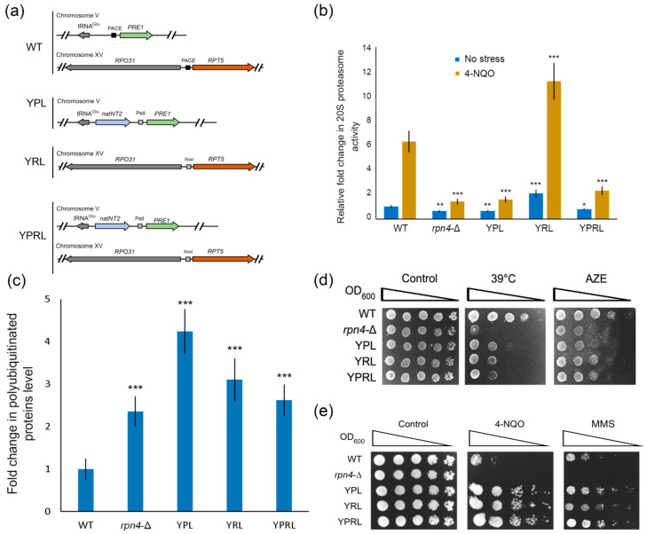
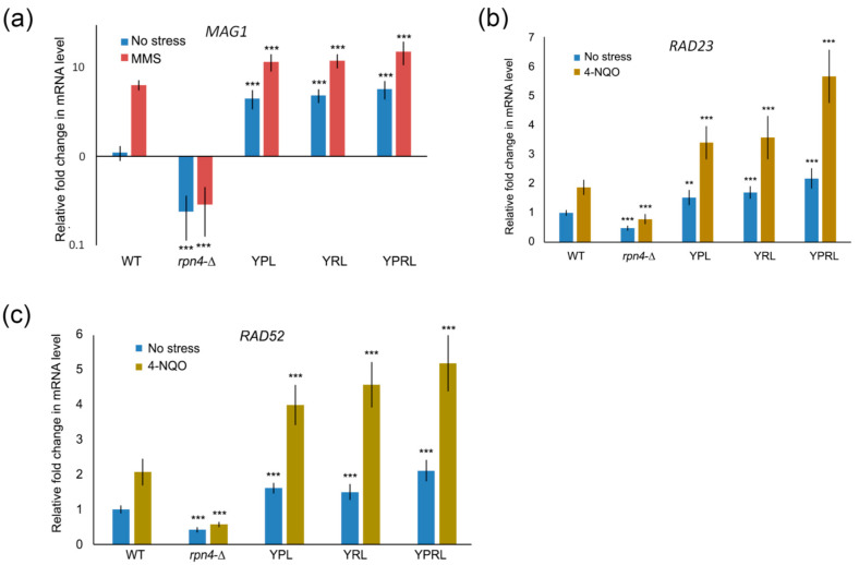
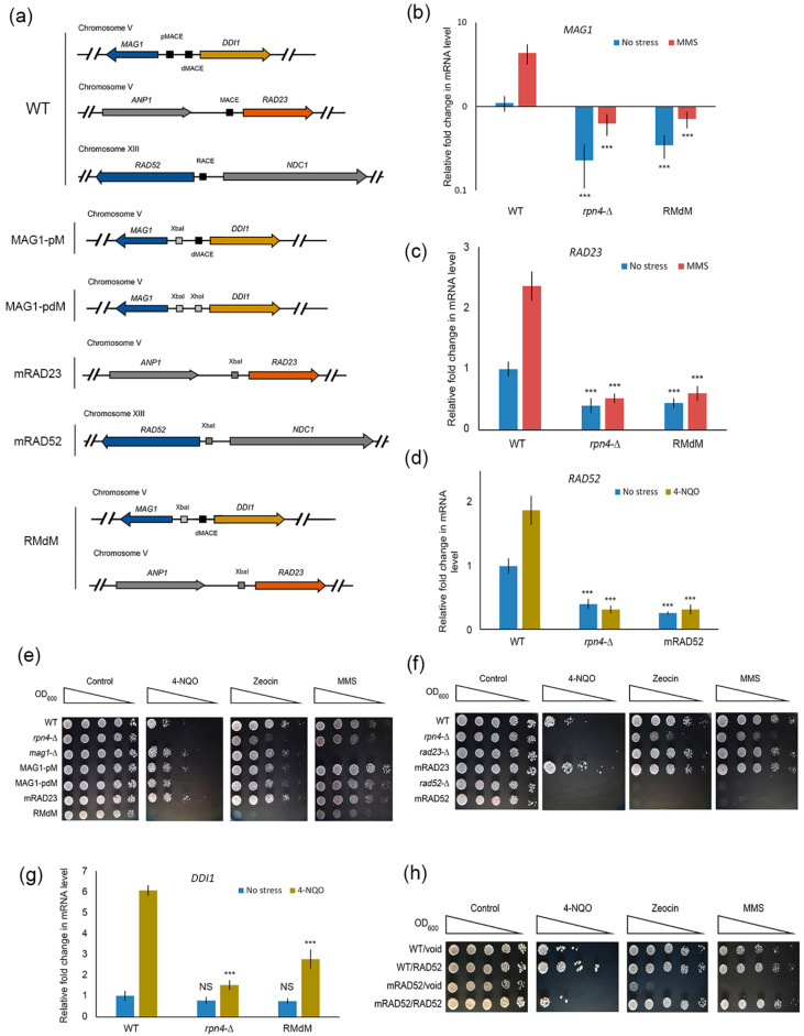
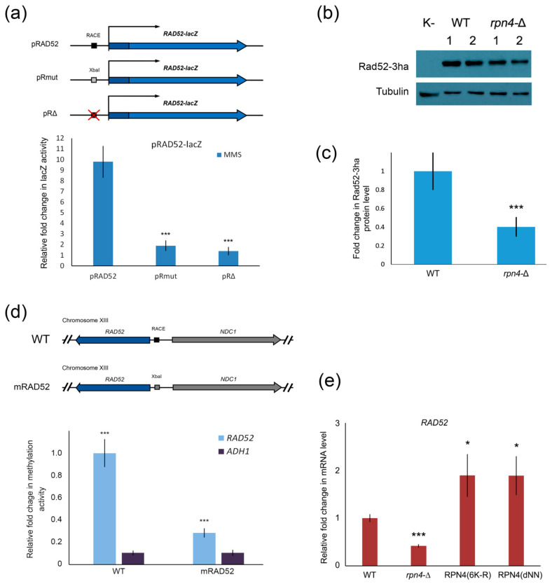
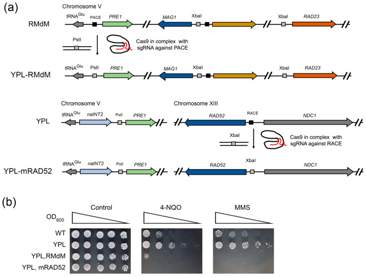
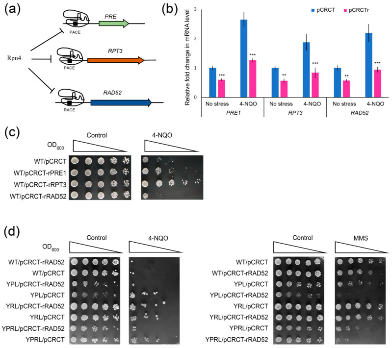
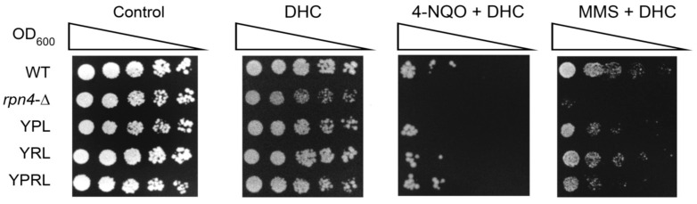
Environmental and intracellular factors often damage DNA, but multiple DNA repair pathways maintain genome integrity. In yeast, the 26S proteasome and its transcriptional regulator and substrate Rpn4 are involved in DNA damage resistance. Paradoxically, while proteasome dysfunction may induce hyper-resistance to DNA-damaging agents, Rpn4 malfunction sensitizes yeasts to these agents. Previously, we proposed that proteasome inhibition causes Rpn4 stabilization followed by the upregulation of Rpn4-dependent DNA repair genes and pathways. Here, we aimed to elucidate the key Rpn4 targets responsible for DNA damage hyper-resistance in proteasome mutants. We impaired the Rpn4-mediated regulation of candidate genes using the CRISPR/Cas9 system and tested the sensitivity of mutant strains to 4-NQO, MMS and zeocin. We found that the separate or simultaneous deregulation of 19S or 20S proteasome subcomplexes induced MAG1, DDI1, RAD23 and RAD52 in an Rpn4-dependent manner. Deregulation of RAD23, DDI1 and RAD52 sensitized yeast to DNA damage. Genetic, epigenetic or dihydrocoumarin-mediated RAD52 repression restored the sensitivity of the proteasome mutants to DNA damage. Our results suggest that the Rpn4-mediated overexpression of DNA repair genes, especially RAD52, defines the DNA damage hyper-resistant phenotype of proteasome mutants. The developed yeast model is useful for characterizing drugs that reverse the DNA damage hyper-resistance phenotypes of cancers.

Keywords: 26S proteasome; CRISPR/Cas9; DDI1; DNA repair; RAD23; RAD52; Rpn4.


Conflict of interest statement

The authors declare no conflict of interest.

Figures

Figure 1
Figure 1
Yeast mutants with deregulated essential proteasomal subunits are hyper-resistant to DNA damage. (a) Schemes of the proteasomal mutant strains. (b) The 20S proteasome activity in proteasome-mutant strains. The 20S proteasome activity was measured in yeast exponential cultures under normal conditions or after 4-NQO treatment at a final concentration 1 µg/mL for 2 h. The relative signal for the wild-type (WT) strain was set to 1. The bar charts show the means (n = 3) ± SDs. Statistical significance: * p between 0.05 and 0.01, ** p between 0.01 and 0.005 and *** p < 0.001, according to Student’s t test. (c) Polyubiquitinated protein levels as quantified by ImageJ. The developed western blot picture is presented in Figure S1. The signal for polyubiquitinated proteins was normalized to the actin signal. The relative signal for the WT strain was set to 1. The values are the means (n = 3) ± SDs. *** p < 0.001, according to Student’s t test. (d) The proteasomal mutant strains were sensitive to proteotoxic conditions. The plates were incubated for 4 days under heat shock conditions or 3 days in the presence of 100 µg/mL L-azetidine-2-carboxylic acid (AZE). (e) The proteasomal mutant strains were hyper-resistant to DNA damage. The plates were incubated for 4 days at 30 °C. 4-NQO was used at a concentration of 0.75 µg/mL. MMS was used at a concentration of 0.017%.
Figure 2
Figure 2
Rpn4-dependent DNA repair genes are upregulated in proteasome-mutant strains. The mRNA expression levels of MAG1 (a), RAD23 (b) and RAD52 (c) were measured by RT-PCR under normal conditions, after 4-NQO treatment at a final concentration of 2 µg/mL for 45 min, or after MMS treatment at a final concentration of 0.2% for 30 min. ACT1 was used as a reference. The relative mRNA level in the wild-type strain under normal conditions was set to 1. The bar charts show the means (n = 3) ± SDs. Statistical significance: ** p between 0.01 and 0.005 and *** p < 0.001, according to Student’s t test.
Figure 3
Figure 3
Deregulation of Rpn4-dependent DNA repair genes sensitizes yeast to DNA damage. (a) Scheme of yeast mutants bearing Rpn4 binding site (MACE or RACE) mutations in the promoter regions of the MAG1-DDI1, RAD23 and RAD52 DNA repair genes. Mutations were introduced into the yeast genome using CRISPR/Cas9-induced template-dependent repair. Both normal and stress-induced expression of MAG1 (b), RAD23 (c) and RAD52 (d) were impaired in strains with mutated Rpn4 binding sites. Exponentially grown cultures were treated with 2 µg/mL 4-NQO for 45 min or 0.2% MMS for 30 min at 30 °C. mRNA levels were measured by RT-PCR and normalized to ACT1. The relative mRNA level in the wild-type (WT) strain under normal conditions was set to 1. The bar charts show the means (n = 3) ± SDs. Statistical significance: *** p < 0.001, according to Student’s t test; (ef) Results of the stress resistance test for mutant strains with deregulated MAG1, RAD23 and RAD52 genes. DNA-damaging agents were used at the following concentrations: 4-NQO, 0.75 µg/mL; MMS, 0.01%; and zeocin, 250 µg/mL. The plates were incubated for 4 days at 30 °C; (g) RT-PCR showed that the mutation of MACE proximal to the MAG1 gene in the MAG1-DDI1 bidirectional promoter decreased the expression of DDI1 under stress conditions. The relative mRNA level in the WT strain under normal conditions was set to 1. The bar charts show the means (n = 3) ± SDs. Statistical significance: NS, non-significant; *** p < 0.001, according to Student’s t test. (h) A plasmid with RAD52 under the control of the native promoter restored resistance to DNA damage in the mRAD52 mutant strain. DNA-damaging agents were used at the following concentrations: 4-NQO, 0.75 µg/mL; MMS, 0.012%; and zeocin, 250 µg/mL. The plates were incubated for 5 days at 30 °C.
Figure 4
Figure 4
Rpn4 regulates RAD52 directly via RACE. (a) RAD52-lacZ translational fusion reporters showed that the RACE element is required for DNA damage-mediated induction of RAD52. Schemes of the RAD52-lacZ reporters used are given. pRAD52 bears the RAD52 promoter and 5′ part of the RAD52 ORF fused in-frame to the lacZ gene. pRmut differs from pRAD52 only by substitution of the 5′-AGTGGC-3′ part of the RACE element with the XbaI site (5′-TCTAGA-3′). The pRΔ construct differs from pRAD52 by deletion of the RACE element. The lacZ activity is reported relative to that of the pRΔ construct. The bar charts show the means (n = 3) ± SDs. Statistical significance: NS, non-significant; *** p < 0.001, according to Student’s t test. (b) Western blot analysis of Rad52-3ha levels in the wild-type (WT) and rpn4-Δ strains. Numbers designate independent colonies. The full image of the developed western blot is presented in Figure S4. (c) Rad52-3ha levels quantified by ImageJ software. The signal for Rad52-3ha was normalized to the tubulin signal. The relative signal for the WT strain was set to 1. The values indicate the means (n = 4) ± SDs. *** p < 0.001, according to Student’s t test. (d) RACE mutation inhibited the Rpn4 interaction with the RAD52 promoter. The methylation signal of the Dam-Rpn4 chimeric reporter protein was normalized to the signal from the mutant reporter Dam-Rpn4(C-A) with impaired Rpn4-binding activity. The ADH1 gene is not an Rpn4 target and was used as a negative control. The relative Dam-Rpn4 signal on the RAD52 promoter in the WT strain was set to 1. The bars and error bars are the means (n = 3) ± SDs. *** p < 0.001, according to Student’s t test. (e) Rpn4 stabilization was sufficient to induce RAD52. The mRNA level of RAD52 in the WT strain expressing stabilized Rpn4 forms was measured using RT-PCR. Rpn4 was stabilized by the deletion of degradation signals (RPN4-dNN) or the mutation of all six N-terminal lysines that contribute to Rpn4 polyubiquitination (Rpn4(6K-R)). The RAD52 mRNA level in the WT strain was set to 1. The values are the means (n = 3) ± SDs. * 0.01 < p < 0.05, *** p < 0.001, according to Student’s t test.
Figure 5
Figure 5
Deregulation of Rpn4-dependent DNA repair genes causes sensitivity to DNA damage in proteasome-mutant strains. (a) Schemes for the creation of mutants with deregulation of both PRE1 and DNA repair genes. Mutations were introduced by CRISPR/Cas9-induced template-dependent repair. (b) The deregulation of DNA repair genes on a YPL background sensitizes mutant yeast to DNA damage. DNA-damaging agents were used at the following concentrations: 4-NQO, 0.7 µg/mL; MMS, 0.016%. The plates were incubated for 4 days at 30 °C.
Figure 6
Figure 6
CRISPR/Cas9-mediated repression of proteasomal and RAD52 genes. (a) Scheme of the experiment. In complex with a short sgRNA, SpyCas9 binds to the PACE or PACE-like element, thereby inhibiting Rpn4 binding. (b) RT-PCR confirmed the CRISPR/Cas9-mediated repression of proteasomal and RAD52 genes in the wild-type (WT) strain both under normal conditions and upon 4-NQO treatment (2 µg/mL for 45 min). The mRNA level of the corresponding gene in the WT strain transformed with the empty pCRCT vector was set to 1. pCRCTr denotes the WT strain transformed with the pCRCT plasmid bearing a short spacer against PACEs in the PRE1 or RPT3 proteasomal genes or RACE in the RAD52 promoter. The values are the means (n = 3) ± SDs. ** 0.05 < p < 0.001, *** p < 0.001, according to Student’s t test. (c) CRISPR/Cas9-mediated repression of the proteasomal gene PRE1 or RPT3 induced hyper-resistance to 4-NQO, while RAD52 repression sensitized yeast to 4-NQO. (d) CRISPR/Cas9-mediated repression of RAD52 sensitized yeast mutants with deregulated proteasomal subunits to DNA-damaging agents. Concentrations of the DNA-damaging agents used: 4-NQO, 0.85 µg/mL; MMS, 0.015%. The plates were incubated for 5 days at 30 °C.
Figure 7
Figure 7
DHC reverses the DNA damage hyper-resistance phenotype of proteasome mutants. Concentrations of chemicals used: DHC, 3 mM; 4-NQO, 0.45 µg/mL; MMS, 0.0175%. The plates were incubated for 4 days at 30 °C.

Similar articles

Cited by

References

    1. Chatterjee N., Walker G.C. Mechanisms of DNA damage, repair, and mutagenesis. Environ. Mol. Mutagen. 2017;58:235–263. doi: 10.1002/em.22087. - DOI - PMC - PubMed
    1. Tubbs A., Nussenzweig A. Endogenous DNA damage as a source of genomic instability in cancer. Cell. 2017;168:644–656. doi: 10.1016/j.cell.2017.01.002. - DOI - PMC - PubMed
    1. Cannan W.J., Pederson D.S. Mechanisms and consequences of double-strand DNA break formation in chromatin. J. Cell. Physiol. 2016;231:3–14. doi: 10.1002/jcp.25048. - DOI - PMC - PubMed
    1. Hoeijmakers J.H. DNA damage, aging, and cancer. N. Engl. J. Med. 2009;361:1475–1485. doi: 10.1056/NEJMra0804615. - DOI - PubMed
    1. Giglia-Mari G., Zotter A., Vermeulen W. DNA damage response. Cold Spring Harb. Perspect. Biol. 2011;3:a000745. doi: 10.1101/cshperspect.a000745. - DOI - PMC - PubMed

MeSH terms

Substances