

Extracellular Vesicles in the Development of the Non-Alcoholic Fatty Liver Disease: An Update
- PMID: 33143043
- PMCID: PMC7693409
- DOI: 10.3390/biom10111494
Extracellular Vesicles in the Development of the Non-Alcoholic Fatty Liver Disease: An Update
Abstract
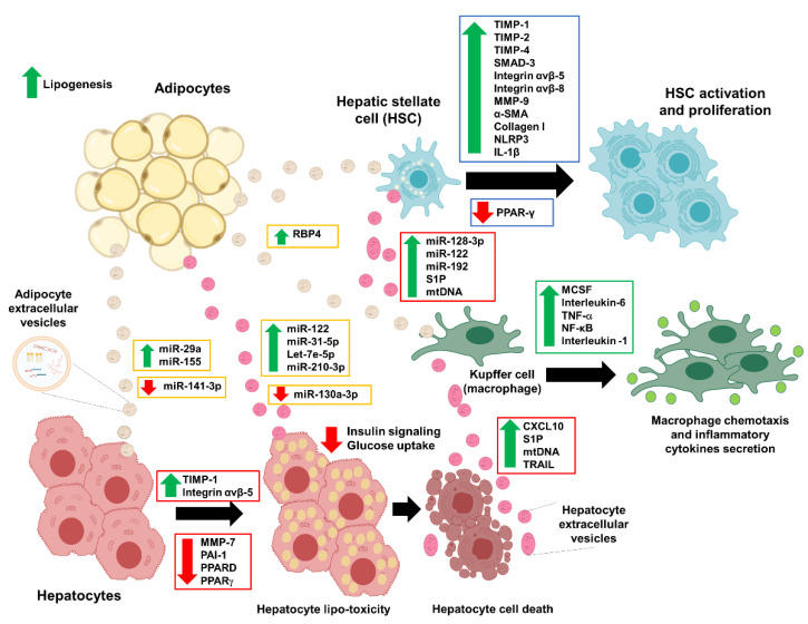
Non-alcoholic fatty liver disease (NAFLD) is a broad spectrum of liver damage disease from a simple fatty liver (steatosis) to more severe liver conditions such as non-alcoholic steatohepatitis (NASH), fibrosis, and cirrhosis. Extracellular vesicles (EVs) are a heterogeneous group of small membrane vesicles released by various cells in normal or diseased conditions. The EVs carry bioactive components in their cargos and can mediate the metabolic changes in recipient cells. In the context of NAFLD, EVs derived from adipocytes are implicated in the development of whole-body insulin resistance (IR), the hepatic IR, and fatty liver (steatosis). Excessive fatty acid accumulation is toxic to the hepatocytes, and this lipotoxicity can induce the release of EVs (hepatocyte-EVs), which can mediate the progression of fibrosis via the activation of nearby macrophages and hepatic stellate cells (HSCs). In this review, we summarized the recent findings of adipocyte- and hepatocyte-EVs on NAFLD disease development and progression. We also discussed previous studies on mesenchymal stem cell (MSC) EVs that have garnered attention due to their effects on preventing liver fibrosis and increasing liver regeneration and proliferation.
Keywords: NAFLD; adipocyte; apoptotic bodies; cell to cell communication; exosome; fibrosis; hepatocyte; insulin resistance; microvesicles.
Conflict of interest statement
The authors declare no conflict of interest. The funders had no role in the design of the study; in the collection, analyses, or interpretation of data; in the writing of the manuscript, or in the decision to publish the results.
Figures
Similar articles
-
Extracellular vesicles derived from fat-laden hepatocytes undergoing chemical hypoxia promote a pro-fibrotic phenotype in hepatic stellate cells.Biochim Biophys Acta Mol Basis Dis. 2020 Oct 1;1866(10):165857. doi: 10.1016/j.bbadis.2020.165857. Epub 2020 Jun 5. Biochim Biophys Acta Mol Basis Dis. 2020. PMID: 32512191
-
Insulin resistance promotes Lysyl Oxidase Like 2 induction and fibrosis accumulation in non-alcoholic fatty liver disease.Clin Sci (Lond). 2017 Jun 7;131(12):1301-1315. doi: 10.1042/CS20170175. Print 2017 Jun 1. Clin Sci (Lond). 2017. PMID: 28468951
-
Lipotoxic hepatocyte derived LIMA1 enriched small extracellular vesicles promote hepatic stellate cells activation via inhibiting mitophagy.Cell Mol Biol Lett. 2024 May 31;29(1):82. doi: 10.1186/s11658-024-00596-4. Cell Mol Biol Lett. 2024. PMID: 38822260 Free PMC article.
-
Extracellular vesicles in fatty liver disease and steatohepatitis: Role as biomarkers and therapeutic targets.Liver Int. 2023 Feb;43(2):292-298. doi: 10.1111/liv.15490. Epub 2022 Dec 30. Liver Int. 2023. PMID: 36462157 Review.
-
Mechanisms of Fibrosis Development in Nonalcoholic Steatohepatitis.Gastroenterology. 2020 May;158(7):1913-1928. doi: 10.1053/j.gastro.2019.11.311. Epub 2020 Feb 8. Gastroenterology. 2020. PMID: 32044315 Free PMC article. Review.
Cited by
-
Nonalcoholic Fatty Liver Disease (NAFLD): Pathogenesis and Noninvasive Diagnosis.Biomedicines. 2021 Dec 22;10(1):15. doi: 10.3390/biomedicines10010015. Biomedicines. 2021. PMID: 35052690 Free PMC article. Review.
-
Extracellular vesicles: mechanisms and prospects in type 2 diabetes and its complications.Front Endocrinol (Lausanne). 2025 Mar 17;15:1521281. doi: 10.3389/fendo.2024.1521281. eCollection 2024. Front Endocrinol (Lausanne). 2025. PMID: 40212823 Free PMC article. Review.
-
The Role of microRNA-22 in Metabolism.Int J Mol Sci. 2025 Jan 17;26(2):782. doi: 10.3390/ijms26020782. Int J Mol Sci. 2025. PMID: 39859495 Free PMC article. Review.
-
Extracellular vesicles-derived ferritin from lipid-induced hepatocytes regulates activation of hepatic stellate cells.Heliyon. 2024 Jun 27;10(13):e33741. doi: 10.1016/j.heliyon.2024.e33741. eCollection 2024 Jul 15. Heliyon. 2024. PMID: 39027492 Free PMC article.
-
The Potential Role of Gut Microbial-Derived Exosomes in Metabolic-Associated Fatty Liver Disease: Implications for Treatment.Front Immunol. 2022 May 11;13:893617. doi: 10.3389/fimmu.2022.893617. eCollection 2022. Front Immunol. 2022. PMID: 35634340 Free PMC article. Review.
References
-
- Araújo A.R., Rosso N., Bedogni G., Tiribelli C., Bellentani S. Global epidemiology of non-alcoholic fatty liver disease/non-alcoholic steatohepatitis: What we need in the future. Liver Int. 2018;38(Suppl. 1):47–51. - PubMed
-
- LaBrecque D.R., Abbas Z., Anania F., Ferenci P., Khan A.G., Goh K.L., Hamid S.S., Isakov V., Lizarzabal M., Peñaranda M.M., et al. World gastroenterology organisation global guidelines: Nonalcoholic fatty liver disease and nonalcoholic steatohepatitis. J. Clin. Gastroenterol. 2014;48:467–473. doi: 10.1097/MCG.0000000000000116. - DOI - PubMed
Publication types
MeSH terms
Grants and funding
LinkOut - more resources
Full Text Sources
Medical
