. 2020 Oct 30;12(11):1232.
doi: 10.3390/v12111232.

New RNA Structural Elements Identified in the Coding Region of the Coxsackie B3 Virus Genome

Affiliations

New RNA Structural Elements Identified in the Coding Region of the Coxsackie B3 Virus Genome

Mariola Dutkiewicz et al. Viruses. .

Abstract

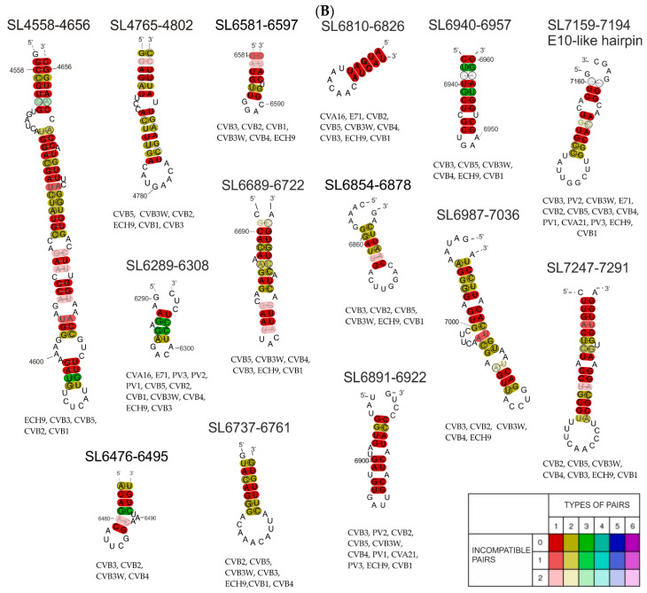
Here we present a set of new structural elements formed within the open reading frame of the virus, which are highly probable, evolutionarily conserved and may interact with host proteins. This work focused on the coding regions of the CVB3 genome (particularly the V4-, V1-, 2C-, and 3D-coding regions), which, with the exception of the cis-acting replication element (CRE), have not yet been subjected to experimental analysis of their structures. The SHAPE technique, chemical modification with DMS and RNA cleavage with Pb2+, were performed in order to characterize the RNA structure. The experimental results were used to improve the computer prediction of the structural models, whereas a phylogenetic analysis was performed to check universality of the newly identified structural elements for twenty CVB3 genomes and 11 other enteroviruses. Some of the RNA motifs turned out to be conserved among different enteroviruses. We also observed that the 3'-terminal region of the genome tends to dimerize in a magnesium concentration-dependent manner. RNA affinity chromatography was used to confirm RNA-protein interactions hypothesized by database searches, leading to the discovery of several interactions, which may be important for virus propagation.

Keywords: CVB3; Coxsackie B3 virus; RNA motif; RNA secondary structure; RNA structural element; RNA structure of coding region; RNA virus; RNA–protein interaction; coxsackievirus B3; enterovirus.


Conflict of interest statement

The authors declare no conflict of interest. The funders had no role in the design of the study; in the collection, analyses, or interpretation of data; in the writing of the manuscript, or in the decision to publish the results.

Figures

Figure 1
Figure 1
Scheme of coxsackievirus B3 genome organization with division into fragments, ca. 750 nt each. Fragments examined in this study are highlighted with colors corresponding to respective regions of the viral genome; UTR—untranslated region; VP1, VP2, VP3, VP4—capsid proteins; 2A, 2B, 2C, 3A, 3B, 3C, 3D—non-structural proteins.
Figure 2
Figure 2
New RNA structural elements identified in the coding part of the Coxsackie B3 virus genome. Most probable secondary structural models of 21 new motifs with experimental mapping results depicted according to the key in the figure. The color of the letters corresponds to the probability values calculated by RNAstructure, according to the key in the figure. Motifs marked in red, orange, and yellow have the highest probability of occurrence in the analyzed RNA fragment and in the corresponding region of the viral genome, so they are considered structurally well-defined. Predicting secondary structure of partly overlapping fragments: F1 and F1–2.
Figure 3
Figure 3
Secondary structure model of F10 RNA, without the last 120 nt of the known pseudoknot. The free energy of the structure is −286.4 kcal/mol. This model displays NMIA modification sites from the SHAPE reaction, DMS modification sites, RNase L digestion, and Pb2+ cleavages, according to the key. In addition, the most interesting structural motifs are marked with their names.
Figure 4
Figure 4
(A) Consensus secondary structures generated by the RNAalifold program for particular RNA motifs identified in the coding part of the Coxsackie B3 virus genome, the “Nancy” strain and 19 other CVB3 strains/isolates. The colors indicate structural conservation according to the key in the figure. (B) Consensus secondary structures generated by RNAalifold program for particular RNA motifs identified in the coding part of the Coxsackie B3 virus genome, the “Nancy” strain and some of 11 other enterovirus strains we investigated. (CVA16-U05876.1|CAU05876 coxsackievirus A16 G-10; CVA21-D00538.1|CXA21CG Human coxsackievirus A21 (strain Coe); CVB1-M16560.1|CXA1G Coxsackievirus B1; CVB2-AF085363.1|Coxsackievirus B2 strain Ohio-1; CVB3-JX312064.1|Human coxsackievirus B3 strain Nancy; CVB3W-U57056.1|CXU57056 Coxsackievirus B3 Woodruff variant; CVB4-X05690.1|Coxsackievirus B4; CVB5-X67706.1|Coxsackievirus B5; PV1-K01392.1|POL3L37 Poliovirus P3/Leon/37 (type 3; PV2-X00595.1|Poliovirus type 2 genome (strain Sabin 2); PV3-V01150.1|Human poliovirus strain Sabin 1; ECH9-X84981.1|Echovirus 9 (strain Hill); E71-AF176044.1|Enterovirus 71.) The colors indicate structural conservation according to the key in the figure.
Figure 4
Figure 4
(A) Consensus secondary structures generated by the RNAalifold program for particular RNA motifs identified in the coding part of the Coxsackie B3 virus genome, the “Nancy” strain and 19 other CVB3 strains/isolates. The colors indicate structural conservation according to the key in the figure. (B) Consensus secondary structures generated by RNAalifold program for particular RNA motifs identified in the coding part of the Coxsackie B3 virus genome, the “Nancy” strain and some of 11 other enterovirus strains we investigated. (CVA16-U05876.1|CAU05876 coxsackievirus A16 G-10; CVA21-D00538.1|CXA21CG Human coxsackievirus A21 (strain Coe); CVB1-M16560.1|CXA1G Coxsackievirus B1; CVB2-AF085363.1|Coxsackievirus B2 strain Ohio-1; CVB3-JX312064.1|Human coxsackievirus B3 strain Nancy; CVB3W-U57056.1|CXU57056 Coxsackievirus B3 Woodruff variant; CVB4-X05690.1|Coxsackievirus B4; CVB5-X67706.1|Coxsackievirus B5; PV1-K01392.1|POL3L37 Poliovirus P3/Leon/37 (type 3; PV2-X00595.1|Poliovirus type 2 genome (strain Sabin 2); PV3-V01150.1|Human poliovirus strain Sabin 1; ECH9-X84981.1|Echovirus 9 (strain Hill); E71-AF176044.1|Enterovirus 71.) The colors indicate structural conservation according to the key in the figure.
Figure 5
Figure 5
Dimerization of the F10 RNA, the 3′-terminal region of the CVB3 genome. (A) Impact of ionic and buffer concentrations; (B) impact of divalent magnesium ions on dimerization process; (C) inhibition of homodimer formation in the presence of “antydimer” (DNA 18-mer), complementary to the F10 in the region of the potential dimerization site; and (D) computer predictions of the region responsible for F10-homodimer formation.
Figure 6
Figure 6
The proteins identified in the RNA-centric affinity chromatography that were found to be interacting with F10 RNA fragment from the 3′-terminus of the CVB3 genome. Possible localizations of the RNA–protein interactions for several proteins is given, based on the presence of a predicted sequence binding site (ATtRACT database) within the F10 sequence. The figure shows the gene names rather than full protein names to minimize the space required. Different colors represent distinct proteins.

Similar articles

Cited by

References

    1. Watts J.M., Dang K.K., Gorelick R.J., Leonard C.W., Bess J.W., Jr., Swanstrom R., Burch C.L., Weeks K.M. Architecture and secondary structure of an entire HIV-1 RNA genome. Nature. 2009;460:711–716. doi: 10.1038/nature08237. - DOI - PMC - PubMed
    1. Li F., Zheng Q., Ryvkin P., Dragomir I., Desai Y., Aiyer S., Valladares O., Yang J., Bambina S., Sabin L.R., et al. Global analysis of RNA secondary structure in two metazoans. Cell Rep. 2012;1:69–82. doi: 10.1016/j.celrep.2011.10.002. - DOI - PubMed
    1. Moss W.N., Steitz J.A. Genome-wide analyses of Epstein-Barr virus reveal conserved RNA structures and a novel stable intronic sequence RNA. BMC Genom. 2013;14:543. doi: 10.1186/1471-2164-14-543. - DOI - PMC - PubMed
    1. Mauger D.M., Golden M., Yamane D., Williford S., Lemon S.M., Martin D.P., Weeks K.M. Functionally conserved architecture of hepatitis C virus RNA genomes. Proc. Natl. Acad. Sci. USA. 2015;112:3692–3697. doi: 10.1073/pnas.1416266112. - DOI - PMC - PubMed
    1. Burrill C.P., Westesson O., Schulte M.B., Strings V.R., Segal M., Andino R. Global RNA structure analysis of poliovirus identifies a conserved RNA structure involved in viral replication and infectivity. J. Virol. 2013;87:11670–11683. doi: 10.1128/JVI.01560-13. - DOI - PMC - PubMed

Publication types

LinkOut - more resources