. 2020 Oct 30;12(11):1236.
doi: 10.3390/v12111236.

Achimota Pararubulavirus 3: A New Bat-Derived Paramyxovirus of the Genus Pararubulavirus

Affiliations

Achimota Pararubulavirus 3: A New Bat-Derived Paramyxovirus of the Genus Pararubulavirus

Kate S Baker et al. Viruses. .

Abstract

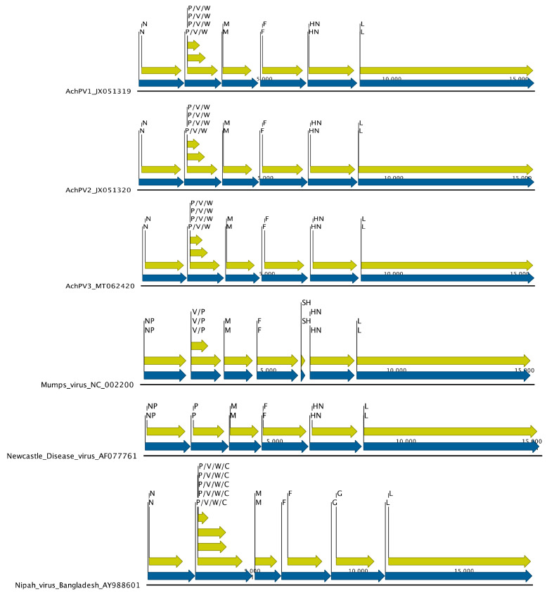
Bats are an important source of viral zoonoses, including paramyxoviruses. The paramyxoviral Pararubulavirus genus contains viruses mostly derived from bats that are common, diverse, distributed throughout the Old World, and known to be zoonotic. Here, we describe a new member of the genus Achimota pararubulavirus 3 (AchPV3) and its isolation from the urine of African straw-coloured fruit bats on primary bat kidneys cells. We sequenced and analysed the genome of AchPV3 relative to other Paramyxoviridae, revealing it to be similar to known pararubulaviruses. Phylogenetic analysis of AchPV3 revealed the failure of molecular detection in the urine sample from which AchPV3 was derived and an attachment protein most closely related with AchPV2-a pararubulavirus known to cause cross-species transmission. Together these findings add to the picture of pararubulaviruses, their sources, and variable zoonotic potential, which is key to our understanding of host restriction and spillover of bat-derived paramyxoviruses. AchPV3 represents a novel candidate zoonosis and an important tool for further study.

Keywords: bat; electron microscopy; genomics; molecular detection; paramyxovirus; pararubulavirus; primary cell lines; virus; virus discovery; zoonosis.


Conflict of interest statement

The authors declare no conflicts of interest.

Figures

Figure 1
Figure 1
Growth of AchPV3 in culture by light (A,B) and electron (C,D) microscopy. Pteropus alecto primary kidney cells uninfected (A) and infected with AchPV3 (B), resulting in multinucleate syncytial cells (black arrows, B). Transmission electron micrographs (C,D) of thin sections reveal cytoplasmic inclusions of viral RNP (white arrow, C). In the negative contrast analysis, pleomorphic particles containing the characteristic Paramyxovirus ribonucleoprotein (RNP) were observed (black arrow, D). Scale bars represent 100 nm.
Figure 2
Figure 2
The phylogenetic relationship of AchPV3 polymerase protein and other paramyxoviruses detected among urine samples using consensus Paramyxovirinae PCR. The phylogenetic tree shows a 176 amino acid alignment of a polymerase protein fragment, and rubulavirus and pararubulavirus fragments detected from bats. Samples starting with U are from Eidolon helvum in Ghana [15]. The AchPV3 sequence is indicated by a black arrow and the scale bars represent the expected number of substitutions per site. Bootstrap values (of 100) of the relevant sites are shown. AchPV1 was detected in, and isolated from, sample U46; AChPV2 was detected in, and isolated from, sample U69; and AchPV3 was isolated from sample U72 but was not detected during PCR screening of the original sample.
Figure 3
Figure 3
Genome architecture of Achimota viruses 1, 2, and 3, Mumps virus, Newcastle Disease Virus, and Nipah virus. Gene boundaries and orientations are indicated by blue arrows and coding sequences by yellow for the nucleoproteins (N/NP), phosphoproteins (P), V/W/C proteins, matrix proteins (M), fusion proteins (F), small hydrophobic protein (SH), attachment proteins (HN/G), and polymerase proteins (L).
Figure 4
Figure 4
The leader and trailer sequences of various pararubulaviruses and the orthorubulaviruses, Porcine orthorubulavirus, and Mumps orthorubulavirus. Those places marked with a dot are where the sequence is identical to Achimota pararubulavirus 1. For viruses where the reverse complementarity of the 5′ trailer (to the 3′ leader) is compromised by an AG couplet, this is indicated by a grey box. AchPV3 is indicated in bold.
Figure 5
Figure 5
The phylogenetic relationships of AchPV3 among the paramyxoviruses. Midpoint-rooted maximum likelihood phylogenetic trees based on a 585 amino acid alignment of the nucleoprotein of Paramyxovirinae members where the pararubulavirus genus is demarcated by a grey circle on the internal node. The AchPV3 sequence is indicated by a black arrow and scale bars represent the expected number of substitutions per site, and the bootstrap values (of 100) of the relevant sites are shown.
Figure 6
Figure 6
The phylogenetic relationship of the AchPV3 Hemagglutinin protein among other paramyxoviruses (pararublaviruses, orthorubulaviruses, and morbilliviruses). Midpoint-rooted maximum likelihood phylogenetic tree based on a 581 amino acid alignment and the scale bar is in expected substitutions per site. Adjacent to the label names is the hexapeptide motif found in the paramyxoviral attachment proteins.

References

    1. Calisher C.H., Childs J.E., Field H.E., Holmes K.V., Schountz T. Bats: Important reservoir hosts of emerging viruses. Clin. Microbiol. Rev. 2006;19:531–545. doi: 10.1128/CMR.00017-06. - DOI - PMC - PubMed
    1. Zhou P., Yang X.L., Wang X.G., Hu B., Zhang L., Zhang W., Si H.R., Zhu Y., Li B., Huang C.L., et al. A pneumonia outbreak associated with a new coronavirus of probable bat origin. Nature. 2020;579:270–273. doi: 10.1038/s41586-020-2012-7. - DOI - PMC - PubMed
    1. Chan J.F., Lau S.K., To K.K., Cheng V.C., Woo P.C., Yuen K.Y. Middle East respiratory syndrome coronavirus: Another zoonotic betacoronavirus causing SARS-like disease. Clin. Microbiol. Rev. 2015;28:465–522. doi: 10.1128/CMR.00102-14. - DOI - PMC - PubMed
    1. Mari Saez A., Weiss S., Nowak K., Lapeyre V., Zimmermann F., Dux A., Kühl H.S., Kaba M., Regnaut S., Merkel K., et al. Investigating the zoonotic origin of the West African Ebola epidemic. EMBO Mol. Med. 2015;7:17–23. doi: 10.15252/emmm.201404792. - DOI - PMC - PubMed
    1. Baker K.S., Leggett R.M., Bexfield N.H., Alston M., Daly G., Todd S., Tachedjian M., Holmes C.E., Crameri S., Wang L.F., et al. Metagenomic study of the viruses of African straw-coloured fruit bats: Detection of a chiropteran poxvirus and isolation of a novel adenovirus. Virology. 2013;441:95–106. doi: 10.1016/j.virol.2013.03.014. - DOI - PMC - PubMed

Publication types

LinkOut - more resources