. 2020 Oct 30;21(21):8130.
doi: 10.3390/ijms21218130.

Processed Eggshell Membrane Powder Is a Promising Biomaterial for Use in Tissue Engineering

Affiliations

Processed Eggshell Membrane Powder Is a Promising Biomaterial for Use in Tissue Engineering

Sissel B Rønning et al. Int J Mol Sci. .

Abstract

The purpose of this study was to investigate the tissue regenerating and biomechanical properties of processed eggshell membrane powder (PEP) for use in 3D-scaffolds. PEP is a low-cost, natural biomaterial with beneficial bioactive properties. Most importantly, this material is available as a by-product of the chicken egg processing (breaking) industry on a large scale, and it could have potential as a low-cost ingredient for therapeutic scaffolds. Scaffolds consisting of collagen alone and collagen combined with PEP were produced and analyzed for their mechanical properties and the growth of primary fibroblasts and skeletal muscle cells. Mechanical testing revealed that a PEP/collagen-based scaffold increased the mechanical hardness of the scaffold compared with a pure collagen scaffold. Scanning electron microscopy (SEM) demonstrated an interconnected porous structure for both scaffolds, and that the PEP was evenly distributed in dense clusters within the scaffold. Fibroblast and skeletal muscle cells attached, were viable and able to proliferate for 1 and 2 weeks in both scaffolds. The cell types retained their phenotypic properties expressing phenotype markers of fibroblasts (TE7, alpha-smooth muscle actin) and skeletal muscle (CD56) visualized by immunostaining. mRNA expression of the skeletal muscle markers myoD, myogenin, and fibroblasts marker (SMA) together with extracellular matrix components supported viable phenotypes and matrix-producing cells in both types of scaffolds. In conclusion, PEP is a promising low-cost, natural biomaterial for use in combination with collagen as a scaffold for 3D-tissue engineering to improve the mechanical properties and promote cellular adhesion and growth of regenerating cells.

Keywords: bovine muscle cells; cell migration; extracellular matrix; human dermal fibroblasts; myofibroblast; processed eggshell membrane powder; scaffold.


Conflict of interest statement

The authors have the following financial competing interests: The author H.P.S., P.B. and R.S. is employed by Biovotec AS, a biotech company aiming to develop wound treatment products. Biovotec has filed a patent on the use of micronized eggshell (WO2016066718: Micronized eggshell membrane particles and the use thereof to promote the healing of wounds). Micronized eggshell membrane, PEP was provided from Biovotec to Nofima for its research activities. Micronized eggshell particles are not commercialized and are today used for research purposes only. This does not alter our adherence to Biomaterials Science policies on sharing data and materials.

Figures
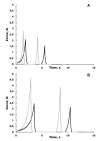
Figure 1
Figure 1
Texture profile curves from two representative collagen scaffolds without (A) and with (B) PEP following a two-cycle compression to 60% of the original scaffold height. The force (N) necessary to maintain a constant speed of the probe was recorded continuously during compression, resulting in a texture profile curve. Black curves—1 h post-swelling, grey curves—4 h post-swelling.
Figure 2
Figure 2
Characterization of scaffolds by SEM-analysis (A,B) and light microscopy with hematoxylin and eosin (HE)-staining (C,D). Scaffold consisting of collagen (A) visualized a porous structure with fibrils interconnected (larger magnification) between sheet-like structures. A similar porous structure was also obtained in scaffold-containing PEP (B), but with aggregates of PEP-structures (larger magnification) within. The fibril-like structures of PEP in a criss-cross pattern was more closely packed. HE-staining demonstrated an organized collagen pattern (C,D), and an even distribution of PEP-clusters of more a pinkish color (D).
Figure 3
Figure 3
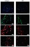
Scaffold with collagen (left panel) and with PEP (right panel) stimulate the growth of fibroblasts. Fibroblasts cells were cultured for one week, and further examined by immunohistochemistry. Expression of fibroblast marker TE7 (light green), proliferation marker KI67 (red), and fibroblast differentiation marker SMA (red). Cell nucleus outlined by Hoechst-staining (blue).
Figure 4
Figure 4
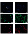
Scaffold with collagen (left panel) and with PEP (right panel) stimulate growth of primary skeletal muscle cells. Primary skeletal muscle cells were cultured for one week, and further examined by immunohistochemistry. Expression of skeletal muscle marker NCAM (light green) localized around nuclear area by Hoechst-staining (blue), together with proliferation marker KI67 (red). Cell nucleus outlined by Hoechst-staining (blue). PEP clusters visualized as diffuse green or a red background.
Figure 5
Figure 5
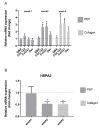
mRNA expression of ECM components and stress markers of fibroblasts cultured in a collagen scaffold with and without PEP. Fibroblasts differentiation and extracellular matrix production (A) is increased together with reduced stress marker HSP (B) in both scaffolds after long-term culturing for two weeks. Bars shows relative mRNA levels presented as fold change (ΔΔCt) in PEP scaffold/collagen scaffold (week 1). Collagen scaffold (week 1) is set to 1 (represented as dotted line). Data are presented as mean ± stdv from three independent cell experiments seeded out in duplicates * Significant difference compared to fibroblast cultured on a collagen scaffold for one week determined by one-way ANOVA using Dunnett’s multiple comparison test.
Figure 6
Figure 6
mRNA expression of ECM components and muscle markers of primary skeletal muscle cells cultured in a collagen scaffold with and without PEP. Skeletal ECM production of collagen (Col1A2) and decorin (Dec) was increased after three weeks of differentiation, whereas the differentiation muscle marker myogenin was reduced. Bars shows relative mRNA levels presented as a fold change (ΔΔCt) in a PEP scaffold/collagen scaffold (week 1). Collagen scaffold (week 1) is set to 1 (represented as dotted line). Data are presented as mean ± stdv from three independent cell experiments seeded out in duplicates. * Significant difference compared to primary skeletal muscle cells cultured on collagen scaffold for one week determined by one-way ANOVA using Dunnett’s multiple comparison test.

References

    1. Smoak M., Mikos A. Advances in biomaterials for skeletal muscle engineering and obstacles still to overcome. Mater. Today Bio. 2020;7:100069. doi: 10.1016/j.mtbio.2020.100069. - DOI - PMC - PubMed
    1. Rodrigues M., Kosaric N., Bonham C.A., Gurtner G.C. Wound Healing: A Cellular Perspective. Physiol. Rev. 2019;99:665–706. doi: 10.1152/physrev.00067.2017. - DOI - PMC - PubMed
    1. Theocharis A.D., Skandalis S.S., Gialeli C., Karamanos N.K. Extracellular matrix structure. Adv. Drug Deliv. Rev. 2016;97:4–27. doi: 10.1016/j.addr.2015.11.001. - DOI - PubMed
    1. Sharma P., Kumar P., Sharma R., Bhatt V.D., Dhot P.S. Tissue Engineering; Current Status & Futuristic Scope. J. Med. Life. 2019;12:225–229. doi: 10.25122/jml-2019-0032. - DOI - PMC - PubMed
    1. Chen G., Ushida T., Tateishi T. Scaffold design for tissue engineering. Macromolec. Biosci. 2002;2:67–77. doi: 10.1002/1616-5195(20020201)2:2<67::AID-MABI67>3.0.CO;2-F. - DOI