. 2020 Oct 30;21(21):8140.
doi: 10.3390/ijms21218140.

How the Pathological Microenvironment Affects the Behavior of Mesenchymal Stem Cells in the Idiopathic Pulmonary Fibrosis

Affiliations

How the Pathological Microenvironment Affects the Behavior of Mesenchymal Stem Cells in the Idiopathic Pulmonary Fibrosis

Martina Bonifazi et al. Int J Mol Sci. .

Abstract

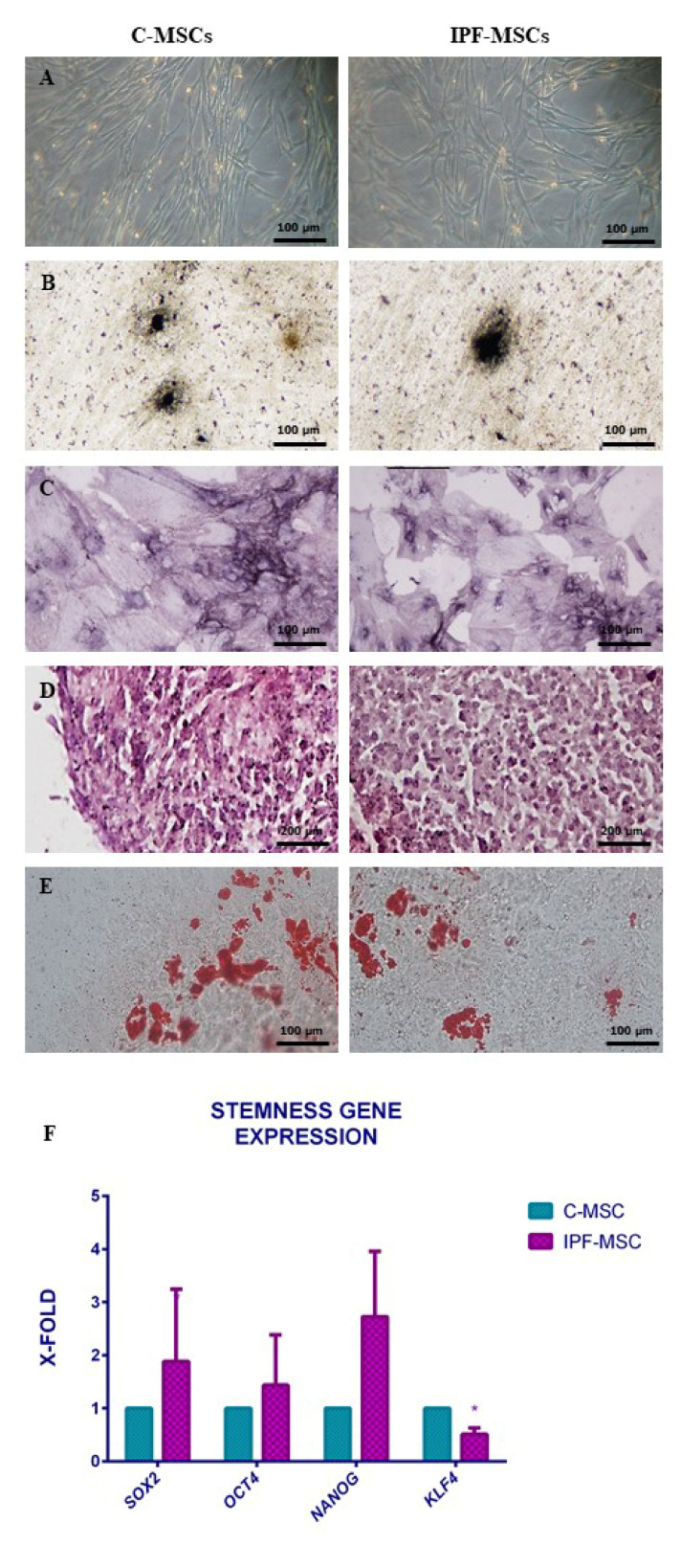
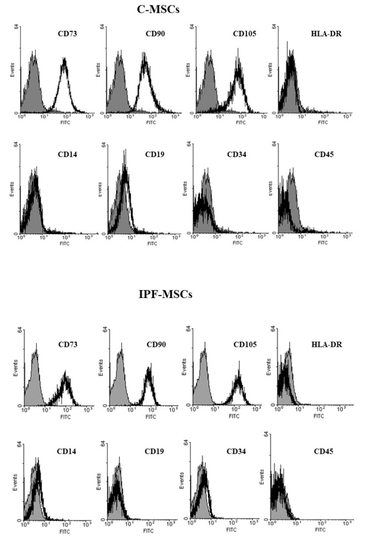
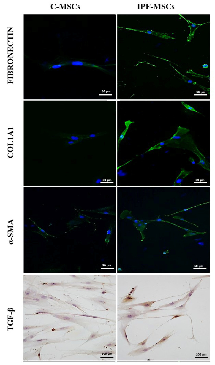
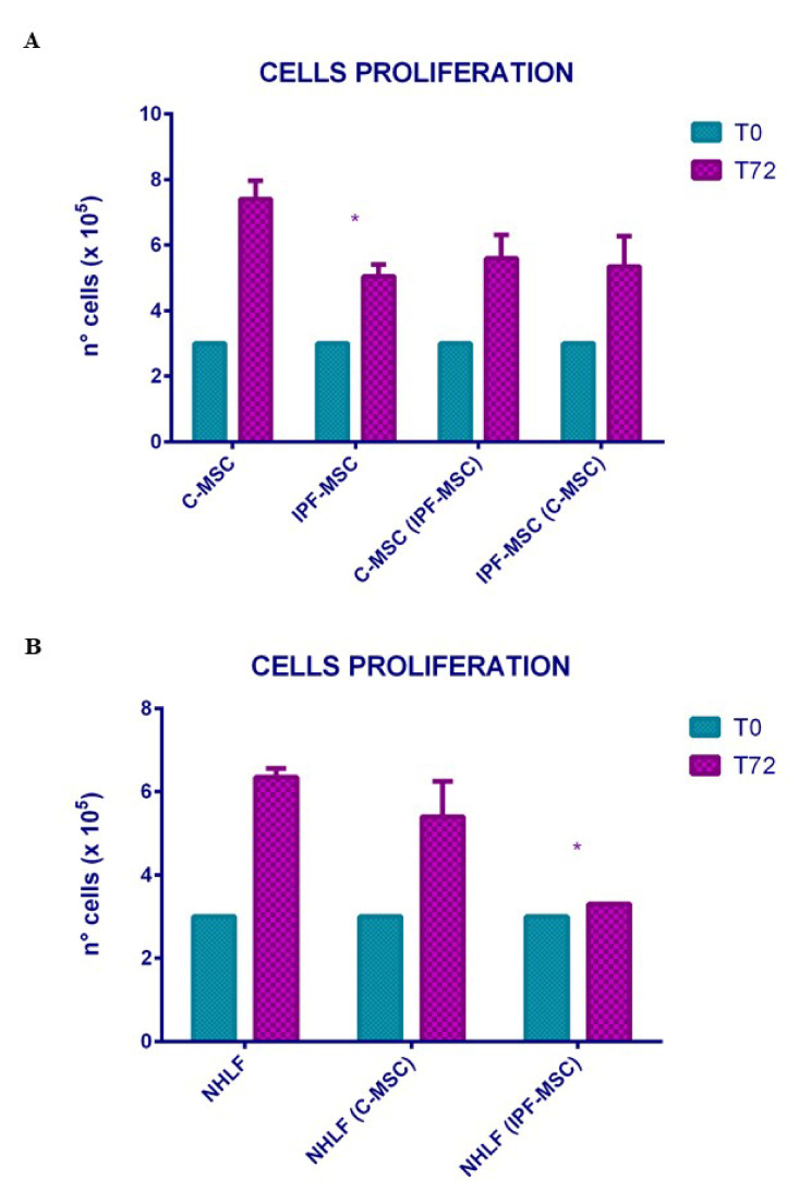
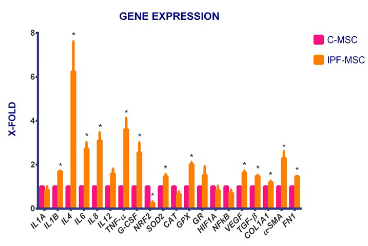
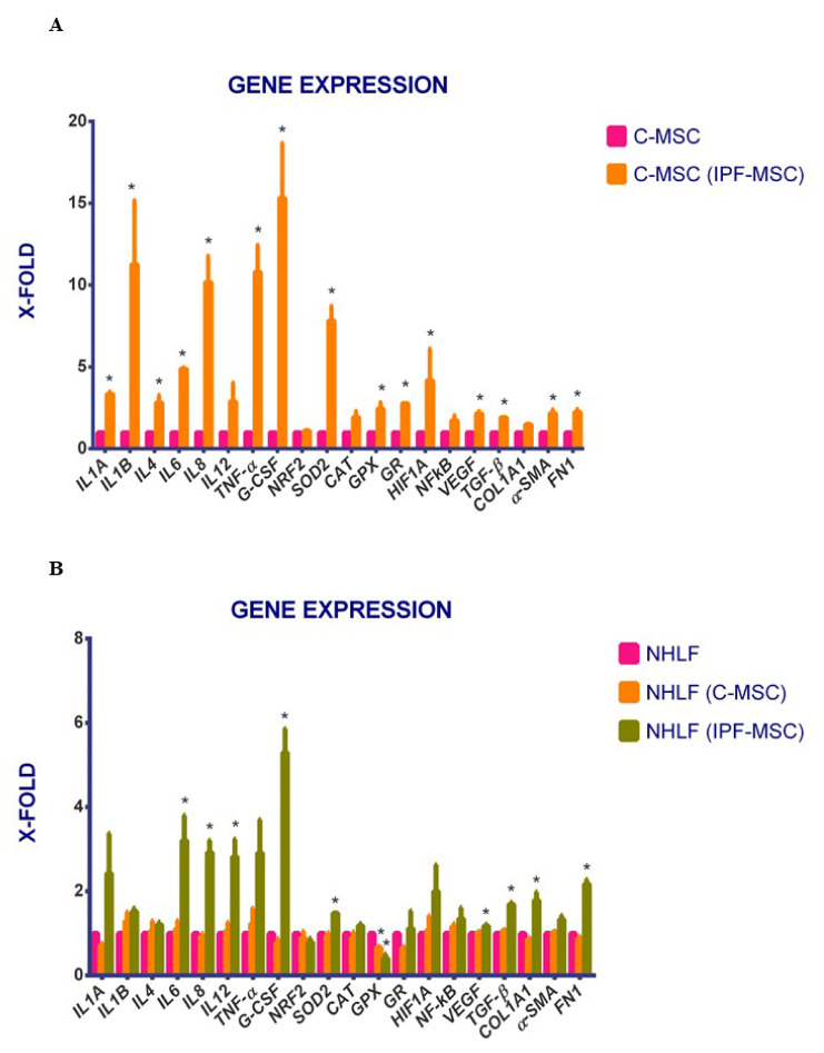
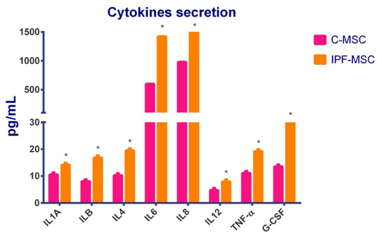
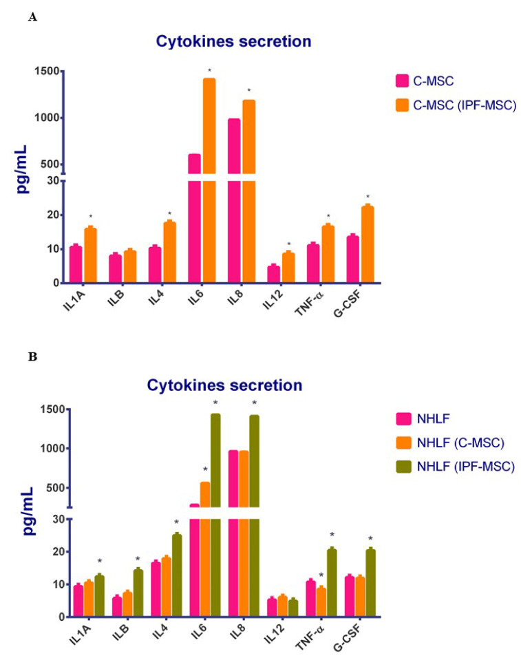
Idiopathic pulmonary fibrosis (IPF) is a chronic disease characterized by fibroblasts activation, ECM accumulation, and diffused alveolar inflammation. The role of inflammation in IPF is still controversial and its involvement may follow nontraditional mechanisms. It is seen that a pathological microenvironment may affect cells, in particular mesenchymal stem cells (MSCs) that may be able to sustain the inflamed microenvironment and influence the surrounding cells. Here MSCs have been isolated from fibrotic (IPF-MSCs) and control (C-MSCs) lung tissue; first cells were characterized and compared by the expression of molecules related to ECM, inflammation, and other interdependent pathways such as hypoxia and oxidative stress. Subsequently, MSCs were co-cultured between them and with NHLF to test the effects of the cellular crosstalk. Results showed that pathological microenvironment modified the features of MSCs: IPF-MSCs, compared to C-MSCs, express higher level of molecules related to ECM, inflammation, oxidative stress, and hypoxia; notably, when co-cultured with C-MSCs and NHLF, IPF-MSCs are able to induce a pathological phenotype on the surrounding cell types. In conclusion, in IPF the pathological microenvironment affects MSCs that in turn can modulate the behavior of other cell types favoring the progression of IPF.

Keywords: idiopathic pulmonary fibrosis; inflammation; mesenchymal stem cells; paracrine effect.


Conflict of interest statement

The authors declare that they have no conflict of interest.

Figures

Figure 1
Figure 1
Mesenchymal stem cells (MSCs) characterization. Phase-contrast images (A) of MSCs derived from control lung tissue (C-MSCs) and from lung of patients affected by idiopathic pulmonary fibrosis (IPF) (IPF-MSCs) after 14 days of culture (scale bar, 100 μm); representative images of osteogenic differentiation assessment by von Kossa staining (B, scale bar 100 μm) and alkaline phosphatase (ALP) reaction (C, scale bar 100 μm); chondrogenic differentiation by safranin-O coloration (D, scale bar 200 μm); adipocyte differentiation by oil red staining (E, scale bar 30 μm). The histogram (F) depicts the expression of genes related to stemness. Levels of expression detected in IPF-MSCs are referred as X-fold with respect to C-MSCs (expressed equal to 1). Data are means ± SD from analyses performed on three separate experiments in triplicates. The * indicates significative differences between C-MSCs and IPF-MSC (unpaired t-test; p < 0.05).
Figure 2
Figure 2
Immunophenotype of Mesenchymal stem Cells (MSCs). Representative FACScan analyses of cell-surface antigen expression, as indicated. Solid gray histograms refer to the negative control (IgG1 isotype control-FITC labeled). No differences were observed between MSCs isolated from the different subgroups.
Figure 3
Figure 3
α-SMA, collagen type 1, fibronectin, and TGF-β1 expression. Indirect immunofluorescence (IIF) analysis of α-SMA, collagen type 1 (COL1A1), and fibronectin and immunocytochemical (ICC) analysis of TGF-β1 performed on mesenchymal stem cells (MSCs) derived from control (C-MSCs) and idiopathic pulmonary fibrosis (IPF-MSCs) tissues. For IIF, a secondary FITC-conjugated antibody was used after incubation with the primary antibodies. Nuclei were counterstained with Hoechst 33342. For ICC of TGF-β1, slides were treated with 3,3-diaminobenzidine and counterstained with Mayer’s hematoxylin.
Figure 4
Figure 4
Effect of co-cultures on cellular proliferation. The histogram (A) indicates the proliferation of mesenchymal stem cells (MSCs) derived from control (C-MSCs) and idiopathic pulmonary fibrosis (IPF-MSCs) tissues (cultured alone) or co-cultured. C-MSCs: MSCs from controls cultured alone; IPF-MSCs: MSCs from patients with IPF cultured alone; C-MSC (IPF-MSCs): C-MSCs after 72 h of co-culture with IPF; IPF-MSCs (C-MSCs): IPF-MSCs after 72 h of co-culture with C-MSCs. The * indicate significative differences referred to C-MSC (one-way ANOVA; p < 0.05). Histogram (B) depicts the proliferation of NHLF cultured alone or co-cultured with C-MSCs (NHLF (C-MSCs)) or with IPF (NHLF (IPF-MSCs)) for 72 h *: p < 0.05 NHLF (IPF-MSCs) vs. NHLF. (The * indicate significative differences referred to C-MSC (two-way ANOVA; p < 0.05).
Figure 5
Figure 5
C-MSCs vs. IPF-MSCs: gene expression. The histogram displays the expression of selected genes referred specifically to inflammation (IL1A, IL1B, IL4, IL6, IL8, IL12, TNF-α, and G-CSF) and to other related pathways such as hypoxia (HIF1A, NFkB, and VEGF), oxidative stress (SOD2, CAT, GPX, GR, and NRF2) and ECM pathway (TGF-β1, COL1A1, α-SMA, and FN1) in C-MSCs and IPF-MSCs. Levels of expression detected in IPF-MSC are referred to as X-fold with respect to C-MSC (express as 1). Data are mean ± SD, over three independent experiments. The * indicates significative differences referred to C-MSC (one-way ANOVA; p < 0.05). C-MSCs: mesenchymal stem cells (MSCs) from controls; IPF-MSCs: MSCs from patients with IPF (idiopathic pulmonary fibrosis).
Figure 6
Figure 6
Gene expression after co-cultures. The histogram displays the expression of selected genes referred specifically to inflammation (IL1A, IL1B, IL4, IL6, IL8, IL12, TNF-α, and G-CSF) and to other related pathways such as hypoxia (HIF1A, NFkB, and VEGF), oxidative stress (SOD2, CAT, GPX, GR, and NRF2) and ECM pathway (TGF-β1, COL1A1, α-SMA, and FN1) after co-culture between C- and IPF-MSC (A). Levels of expression detected in C-MSC(IPF-MSC) is referred to as X-fold with respect to C-MSC (express as 1). The figure (B) shows the expression of the selected genes on NHLF before and after co-culturing with C-and IPF-MSCs. Levels of expression detected in NHLF(C-MSC) and in NHLF(IPF-MSC) are referred to as X-fold with respect to NHLF (express as 1). Data are mean ± SD, over three independent experiments. The * indicate significative differences referred to as C-MSC (one-way ANOVA; p < 0.05). C-MSCs: mesenchymal stem cells (MSCs) from controls cultured alone; C-MSC (IPF-MSCs): C-MSCs after 72 h of co-culture with IPF; NHLF: NHLF cultured alone; NHLF (C-MSCs): NHLF co-cultured with C-MSCs for 72 h; NHLF (IPF-MSCs): NHLF co-cultured with IPF-MSCs for 72 h.
Figure 7
Figure 7
C-MSCs vs. IPF-MSCs: cytokines secretion. The secretion of IL1A, IL1B, IL4, IL6, IL8, IL12, TNF-α, and G-CSF was evaluated by ELISA. Levels of secretion were measured on collected medium after 72 h of culture and are referred to as pg/mL. Data are means ± SD from analyses performed on three separate experiments in triplicates. The * indicate significative differences between C-MSCs and IPF-MSC; (one-way ANOVA; p < 0.05). C-MSCs: mesenchymal stem cells (MSCs) from controls; IPF-MSCs: MSCs from patients with IPF (idiopathic pulmonary fibrosis).
Figure 8
Figure 8
Cytokines secretion after co-cultures. The secretion of IL1A, IL1B, IL4, IL6, IL8, IL12, TNF-α, and G-CSF was evaluated by ELISA after co-culture between C- and IPF-MSC (A) and among NHLF and MSCs (B). Levels of secretion were measured on collected medium after 72 h of co-culture and are referred to as pg/mL. Data are means ± SD from analyses performed on three separate experiments in triplicates. In figure (A) the * indicates significative differences between C-MSCs and C-MSC(IPF-MSC); in figure (B) the * indicates significative differences between NHLF and NHLF(C-MSC) and between NHLF and NHLF(IPF.MSC); (one-way ANOVA; p < 0.05). C-MSCs: mesenchymal stem cells (MSCs) from controls cultured alone; C-MSC (IPF-MSCs): C-MSCs after 72 h of co-culture with IPF; NHLF: NHLF cultured alone; NHLF (C-MSCs): NHLF co-cultured with C-MSCs for 72 h; NHLF (IPF-MSCs): NHLF co-cultured with IPF-MSCs for 72 h.

Similar articles

Cited by

References

    1. Liu Y.M., Nepali K., Liou J.P. Idiopathic pulmonary fibrosis: Current status, recent progress, and emerging targets. J. Med. Chem. 2017;60:527–553. doi: 10.1021/acs.jmedchem.6b00935. - DOI - PubMed
    1. Padilla M. Idiopathic pulmonary fibrosis: The role of pathobiology in making a definitive diagnosis. Am. J. Manag. Care. 2015;21:s276–s283. - PubMed
    1. King T.E., Jr., Tooze J.A., Schwarz M.I., Brown K.R., Cherniack R.M. Predicting survival in idiopathic pulmonary fibrosis: Scoring system and survival model. Am. J. Respir. Crit. Care Med. 2001;164:1171–1181. doi: 10.1164/ajrccm.164.7.2003140. - DOI - PubMed
    1. Tian Y., Li H., Gao Y., Liu C., Qiu T., Wu H., Cao M., Zhang Y., Ding H., Chen J., et al. Quantitative proteomic characterization of lung tissue in idiopathic pulmonary fibrosis. Clin. Proteom. 2019;16:1–11. doi: 10.1186/s12014-019-9226-4. - DOI - PMC - PubMed
    1. Steele M.P., Schwartz D.A. Molecular mechanisms in progressive idiopathic pulmonary fibrosis. Annu. Rev. Med. 2013;64:265–276. doi: 10.1146/annurev-med-042711-142004. - DOI - PubMed

MeSH terms

LinkOut - more resources