. 2020 Oct 30;21(21):8142.
doi: 10.3390/ijms21218142.

Age-Dependent and Sleep/Seizure-Induced Pathomechanisms of Autosomal Dominant Sleep-Related Hypermotor Epilepsy

Affiliations

Age-Dependent and Sleep/Seizure-Induced Pathomechanisms of Autosomal Dominant Sleep-Related Hypermotor Epilepsy

Kouji Fukuyama et al. Int J Mol Sci. .

Abstract

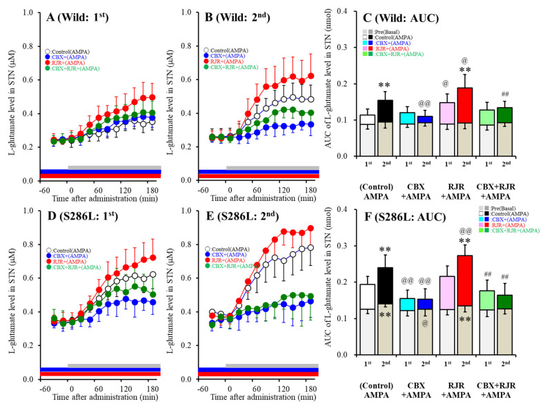
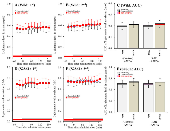
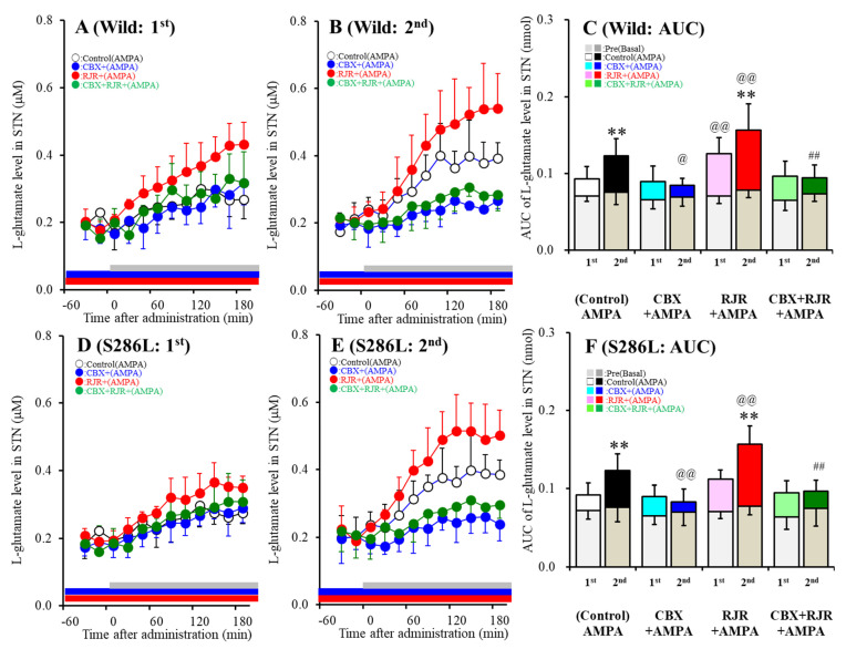
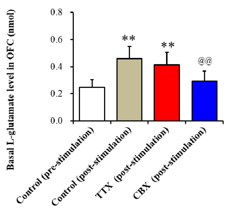
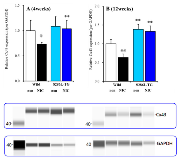
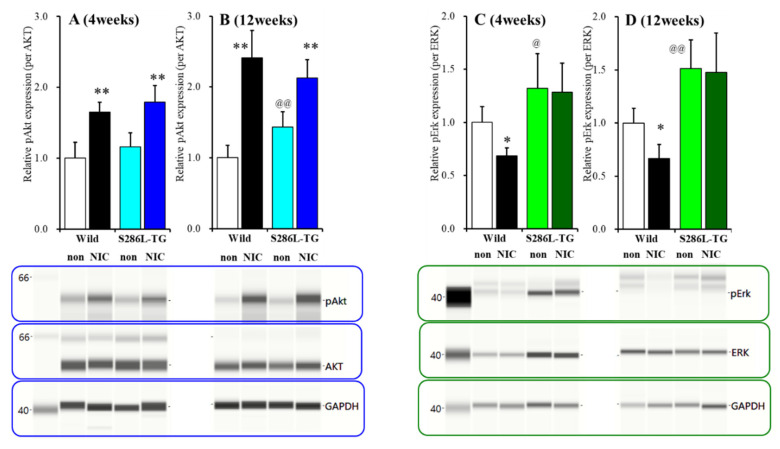
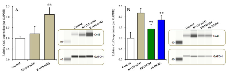
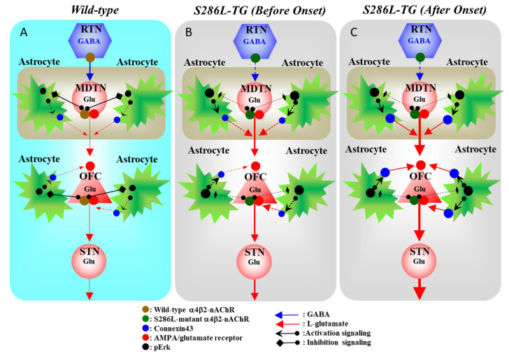
The loss-of-function S284L-mutant α4 subunit of the nicotinic acetylcholine receptor (nAChR) is considered to contribute to the pathomechanism of autosomal dominant sleep-related hypermotor epilepsy (ADSHE); however, the age-dependent and sleep-related pathomechanisms of ADSHE remain to be clarified. To explore the age-dependent and sleep-induced pathomechanism of ADSHE, the present study determined the glutamatergic transmission abnormalities associated with α4β2-nAChR and the astroglial hemichannel in the hyperdirect and corticostriatal pathways of ADSHE model transgenic rats (S286L-TG) bearing the rat S286L-mutant Chrna4 gene corresponding to the human S284L-mutant CHRNA4 gene of ADSHE, using multiprobe microdialysis and capillary immunoblotting analyses. This study could not detect glutamatergic transmission in the corticostriatal pathway from the orbitofrontal cortex (OFC) to the striatum. Before ADSHE onset (four weeks of age), functional abnormalities of glutamatergic transmission compared to the wild-type in the cortical hyperdirect pathway, from OFC to the subthalamic nucleus (STN) in S286L-TG, could not be detected. Conversely, after ADSHE onset (eight weeks of age), glutamatergic transmission in the hyperdirect pathway of S286L-TG was enhanced compared to the wild-type. Notably, enhanced glutamatergic transmission of S286L-TG was revealed by hemichannel activation in the OFC. Expression of connexin43 (Cx43) in the OFC of S286L-TG was upregulated after ADSHE onset but was almost equal to the wild-type prior to ADSHE onset. Differences in the expression of phosphorylated protein kinase B (pAkt) before ADSHE onset between the wild-type and S286L-TG were not observed; however, after ADSHE onset, pAkt was upregulated in S286L-TG. Conversely, the expression of phosphorylated extracellular signal-regulated kinase (pErk) was already upregulated before ADSHE onset compared to the wild-type. Both before and after ADSHE onset, subchronic nicotine administration decreased and did not affect the both expression of Cx43 and pErk of respective wild-type and S286L-TG, whereas the pAkt expression of both the wild-type and S286L-TG was increased by nicotine. Cx43 expression in the plasma membrane of the primary cultured astrocytes of the wild-type was increased by elevation of the extracellular K+ level (higher than 10 mM), and the increase in Cx43 expression in the plasma membrane required pErk functions. These observations indicate that a combination of functional abnormalities, GABAergic disinhibition, and upregulated pErk induced by the loss-of-function S286L-mutant α4β2-nAChR contribute to the age-dependent and sleep-induced pathomechanism of ADSHE via the upregulation/hyperactivation of the Cx43 hemichannels.

Keywords: L-glutamate; autosomal dominant sleep-related hypermotor epilepsy; basal ganglia; extracellular signal-regulated kinase; hemichannel; protein kinase B.


Conflict of interest statement

The authors state no conflict of interest.

Figures

Figure 1
Figure 1
Effects of the local administration of 100 μM RJR2403 (selective α4β2-nAChR agonist) and 100 μM carbenoxolone (CBX; hemichannel inhibitor) into the orbitofrontal cortex (OFC) on 100 μM amino-3-(3-hydroxy-5-methyl-isoxazol-4-yl)propanoic acid (AMPA)-evoked (perfusion with 100 μM AMPA into the OFC) L-glutamate release in the subthalamic nucleus (STN), before (A,D) and after (B,E) hemichannel activation (FCHK-MRS (Ca2+-free with 100 mM K+ containing modified Ringer’s solution) perfusion) of the wild-type (A,B) and S286L-TG (D,E). The perfusion medium in the OFC began with MRS, with or without (control) 100 μM CBX (blue bars) or 100 μM RJR2403 (red bars). After stabilization of the L-glutamate level in the STN, the perfusion medium was switched to the same MRS containing 100 μM AMPA, for 180 min (first AMPA-evoked stimulation: gray bars). After stabilization of the L-glutamate level in the STN, the perfusion medium was switched from MRS to FCHK-MRS, for 20 min (hemichannel activation). After stabilization of the L-glutamate level in the STN, the perfusion medium was again switched to the same MRS containing 100 μM AMPA, for 180 min (second AMPA-evoked stimulation: gray bars). Ordinates (A,B,D,E) indicate the mean extracellular L-glutamate level (μM) (n = 6), and abscissas indicate the time after AMPA-evoked stimulations (min). (C,F) indicate the area under the curve (AUC) value of the extracellular L-glutamate level (nmol) before (basal extracellular L-glutamate level) and during perfusion with AMPA (from 20 to 180 min) of the wild-type (A,B) and S286L-TG (D,E). Notably, the gray columns in (C,F) indicate the AUC values of the basal extracellular levels of L-glutamate before AMPA-evoked stimulation (basal L-glutamate release) (over −60 to 0 min in (A,B,D,E)). * p < 0.05, ** p < 0.01; relative to the first (first AMPA-evoked stimulation), @p < 0.05, @@p < 0.01; relative to the control, # p < 0.05, ## p < 0.01; relative to RJR by MANOVA with Tukey’s multiple comparison. The F-values of the L-glutamate level in the STN, according to a multivariate analysis of variance (MANOVA), were Fevent(1,80) = 15.1(p < 0.01), FRJR(1,80) = 17.3(p < 0.01), FCBX(1,80) = 76.8(p < 0.01), Fgenotype(1,80) = 128.6 (p < 0.01), Fevent*RJR(1,80) = 0.2 (p > 0.05), Fevent*CBX(1,80) = 21.3(p < 0.01), Fevent*genotype(1,80) = 0.1(p > 0.05), FRJR*CBX(1,80) = 1.8(p > 0.05), FRJR*genotype(1,80)= 0.1(p > 0.05), FCBX*genotype(1,80) = 13.9(p < 0.01), Fevent*RJR*CBX(1,80) = 0.1(p > 0.05), Fevent*RJR*genotype(1,80) = 0.1 (p > 0.05), Fevent*CBX*genotype(1,80) = 0.6(p > 0.05), FRJR*CBX*genotype(1,80) = 0.1(p > 0.05), and Fevent*RJR*CBX*genotype (1,80)= 0.7 (p > 0.05).
Figure 2
Figure 2
Effects of the local administration of 100 μM RJR2403 into the OFC on 100 μM AMPA-evoked (perfusion with 100 μM AMPA into the OFC) L-glutamate release in the striatum, before (A,D) and after (B,E) hemichannel activation (FCHK-MRS perfusion for 20 min) of the wild-type (A,B) and S286L-TG (D,E). The perfusion medium in the OFC began with MRS with or without (control) 100 μM RJR2403 (red bars). After stabilization of the L-glutamate level in the striatum, the perfusion medium was switched to the same MRS containing 100 μM AMPA for 180 min (first AMPA-evoked stimulation: gray bars). After stabilization of the L-glutamate level in the striatum, the perfusion medium was switched from MRS to FCHK-MRS for 20 min (hemichannel activation). After stabilization of the L-glutamate level in the striatum, the perfusion medium was again switched to the same MRS containing 100 μM AMPA for 180 min (second AMPA-evoked stimulation: gray bars). Ordinates (A,B,D,E) indicate the mean extracellular L-glutamate level (μM) (n = 6), and abscissas indicate the time after AMPA-evoked stimulations (min). (C,F) indicate the AUC of the extracellular L-glutamate level (nmol) before (basal extracellular L-glutamate release) and during perfusion with AMPA (from 20 to 180 min) of the wild-type (A,B) and S286L-TG (D,E), respectively. Notably, the gray columns in (C,F) indicate the AUC values of the basal extracellular levels of L-glutamate before AMPA-evoked stimulation (basal L-glutamate release) (over −60 to 0 min in (A,B,D,E)). The F-values of the L-glutamate level in the striatum by MANOVA were Fevent(1,40) = 3.8(p > 0.05), FRJR(1,40) = 0.1(p > 0.05), Fgenotype(1,40) = 40.1(p < 0.01), Fevent*RJR(1,40) = 0.1(p > 0.05), Fevent*genotype(1,40) = 0.1 (p > 0.05), FRJR*genotype(1,40) = 0.1 (p > 0.05), FRJR*genotype(1,40) = 0.1(p > 0.05), and Fevent*RJR*genotype (1,40) = 0.1 (p > 0.05).
Figure 3
Figure 3
Effects of the local administration of 100 μM RJR2403 (selective α4β2-nAChR agonist) and 100 μM CBX (hemichannel inhibitor) into the OFC on 100 μM AMPA-evoked (perfusion with 100 μM AMPA into the OFC) L-glutamate release in the STN, before (A,D) and after (B,E) hemichannel activation (FCHK-MRS (Ca2+-free with 100 mM K+) perfusion) of the wild-type (A,B) and S286L-TG (D,E). Ordinates (A,B,D,E) indicate the mean extracellular L-glutamate level (μM) (n=6), and abscissas indicate the time after AMPA-evoked stimulations (min). (C,F) indicate the AUC value of the extracellular L-glutamate level (nmol) before (basal extracellular L-glutamate level) and during perfusion with AMPA (from 20 to 180 min) for the wild-type (A,B) and S286L-TG (D,E), respectively. Especially, gray columns in (C,F) indicate the AUC values of the basal extracellular levels of L-glutamate before AMPA-evoked stimulation (basal L-glutamate release) (during −60 to 0 min in A,B,D,E). * p < 0.05, ** p < 0.01; relative to the first (first AMPA-evoked stimulation), @p < 0.05, @@p < 0.01; relative to the control, # p < 0.05, ## p < 0.01; relative to RJR by MANOVA with Tukey’s multiple comparison. The F-values of the L-glutamate level in the STN, according to a multivariate analysis of variance (MANOVA), were Fevent(1,80) = 15.1 (p < 0.01), FRJR(1,80) = 21.9 (p < 0.01), FCBX(1,80) = 59.9 (p < 0.01), Fgenotype(1,80) = 0.2 (p > 0.05), Fevent*RJR(1,80) = 0.7 (p > 0.05), Fevent*CBX(1,80) = 19.3 (p < 0.01), Fevent*genotype(1,80) = 0.3 (p > 0.05), FRJR*CBX(1,80) = 6.6 (p < 0.05), FRJR*genotype(1,80) = 0.1 (p > 0.05), FCBX*genotype(1,80) = 0.1 (p > 0.05), Fevent*RJR*CBX(1,80) = 0.1 (p > 0.05), Fevent*RJR*genotype(1,80) = 0.4 (p > 0.05), Fevent*CBX*genotype(1,80) = 0.1 (p > 0.05), FRJR*CBX*genotype(1,80) = 0.2 (p > 0.05), and Fevent*RJR*CBX*genotype(1,80)= 0.1 (p > 0.05).
Figure 4
Figure 4
Effects of the local administration of 1 μM tetrodotoxin (TTX) and 100 μM CBX into the OFC on basal L-glutamate release in the OFC of S286L-TG after FCHK-evoked stimulation. The perfusion medium in the OFC was switched to MRS, with or without (control: post-stimulation: gray column) 1 μM TTX (red column) or 100 μM CBX (blue column). The ordinate AUC of the extracellular L-glutamate level (nmol) for 60 min during perfusion of MRS with or without TTX or CBX is shown. ** p < 0.01, relative to the control (pre-stimulation), and @@ p < 0.01, relative to the control (post-stimulation) by a one-way analysis of variance (ANOVA) with Tukey’s multiple comparison. The F-values of the L-glutamate level in the OFC, according to the one-way ANOVA, are F(3,20) = 9.6 (p < 0.01).
Figure 5
Figure 5
Effects of subchronic nicotine administration on connexin43 (Cx43) expression in the OFC. Effects of systemic subchronic administration of nicotine (50 mg/kg/day for seven days) on Cx43 expression in the OFC plasma membrane fraction before four weeks of age (A), and after 12 weeks of age (B), autosomal dominant sleep-related hypermotor epilepsy (ADSHE) onset of the wild-type and S286L-TG and pseudo-gel images from the capillary immunoblotting results, using anti-GAPDH and anti-connexin43 antibodies for blotting of the plasma membrane fractions. Ordinate: mean ± SD (n = 6) of the relative protein level of Cx43. ** p < 0.01 vs. the wild-type, and @ p < 0.05, @@ p < 0.01 vs. nicotine-free (non) based on a two-way ANOVA with Tukey’s multiple comparison.
Figure 6
Figure 6
Effects of subchronic nicotine administration on the expression of phosphorylated protein kinase B (pAkt) and phosphorylated extracellular signal-regulated kinase (pErk) in the plasma membrane fraction of OFC. Effects of the systemic subchronic administration of nicotine (50 mg/kg/day for seven days) on pAkt and pErk expression in the OFC plasma membrane fraction before four week of age (A,C) and after 12 week of age (B,D), ADSHE onset of the wild-type and S286L-TG and pseudo-gel images, using capillary immunoblotting. Ordinate: mean ± SD (n = 6) of the relative protein level of pErk and pAkt. * p < 0.05, ** p < 0.01 vs. wild-type, and @ p < 0.05, @@ p < 0.01 vs. nicotine-free (non) by two-way ANOVA with Tukey’s multiple comparison.
Figure 7
Figure 7
Effects of subacute administration of an increase in the extracellular K+ level on Cx43 expression in the plasma membrane fraction of primary cultured astrocytes (A). Effects of the inhibitor of Erk (FR180204) and Akt (10-DEBC) on K+-dependent Cx43 expression in the plasma membrane (B). Ordinate: mean ± SD (n = 6) of the relative protein level of Cx43. Concentration-dependent effects of extracellular K+ on Cx43 expression in the plasma membrane fraction of the primary cultured astrocytes were analyzed by a one-way ANOVA (@@ p < 0.01 vs. control). The effects of 20 μM FR180204 and 10 μM 10-DEBC on Cx43 expression in the plasma membrane fraction were analyzed by a Student’s t-test (** p < 0.01 vs. 10 mM K+).
Figure 8
Figure 8
Proposed hypothesis of the multi-stage pathomechanisms of S286L-TG. Proposed hypothesis for the functional abnormalities of glutamatergic transmission in the thalamocortical and cortical hyperdirect pathways in the wild-type (A), S286L-TG before ADSHE onset (B), and after ADSHE onset (C). Reticular thalamic nucleus (RTN) mainly projects GABAergic terminals to various thalamic nuclei, including mediodorsal thalamic nucleus (MDTN). The activation of α4β2-nAChR in the RTN enhances GABAergic transmission in the RTN–MDTN pathways of the wild-type (A), whereas the S286L-mutant α4β2-nAChR impairs the activation of GABAergic transmission in the RTN–MDTN in S286L-TG (B,C). MDTN project glutamatergic terminals to the OFC. In the MDTN, both α4β2-nAChR and the AMPA/glutamate receptor activate glutamatergic transmission to the OFC (AC). Wild-type α4β2-nAChR inhibits astroglial Erk, resulting in the suppression of connexin43 expression in the astroglial plasma membrane (A). Contrary to the wild-type, in S286L-TG, the loss-of-function S286L-mutant α4β2-nAChR lacks suppressive effects on pErk (B,C) but is insufficient to upregulate connexin43 (B). A combination of the persistent/repetitive propagation of the hyperactivation of glutamatergic transmission in MDTN-OFC induced by the GABAergic disinhibition of S286L-TG and pErk upregulation enhances connexin43 expression.

Similar articles

Cited by

References

    1. Scheffer I.E., Bhatia K.P., Lopes-Cendes I., Fish D.R., Marsden C.D., Andermann F., Andermann E., Desbiens R., Cendes F., Manson J.I., et al. Autosomal dominant frontal epilepsy misdiagnosed as sleep disorder. Lancet. 1994;343:515–517. doi: 10.1016/S0140-6736(94)91463-X. - DOI - PubMed
    1. Tinuper P., Bisulli F., Cross J.H., Hesdorffer D., Kahane P., Nobili L., Provini F., Scheffer I.E., Tassi L., Vignatelli L., et al. Definition and diagnostic criteria of sleep-related hypermotor epilepsy. Neurology. 2016;86:1834–1842. doi: 10.1212/WNL.0000000000002666. - DOI - PMC - PubMed
    1. Hebbar M., Mefford H.C. Recent advances in epilepsy genomics and genetic testing. F1000 Res. 2020;9:185. doi: 10.12688/f1000research.21366.1. - DOI - PMC - PubMed
    1. Okada M., Zhu G., Yoshida S., Kaneko S. Validation criteria for genetic animal models of epilepsy. Epilepsy Seizure. 2010;3:109–120. doi: 10.3805/eands.3.109. - DOI
    1. Nobili L., Proserpio P., Combi R., Provini F., Plazzi G., Bisulli F., Tassi L., Tinuper P. Nocturnal frontal lobe epilepsy. Curr. Neurol. Neurosci. Rep. 2014;14:424. doi: 10.1007/s11910-013-0424-6. - DOI - PubMed