

Staged reconstruction of a chronically infected large skull defect using free tissue transfer and a patient-specific polyetheretherketone implant
- PMID: 33143400
- PMCID: PMC7644347
- DOI: 10.7181/acfs.2020.00311
Staged reconstruction of a chronically infected large skull defect using free tissue transfer and a patient-specific polyetheretherketone implant
Abstract
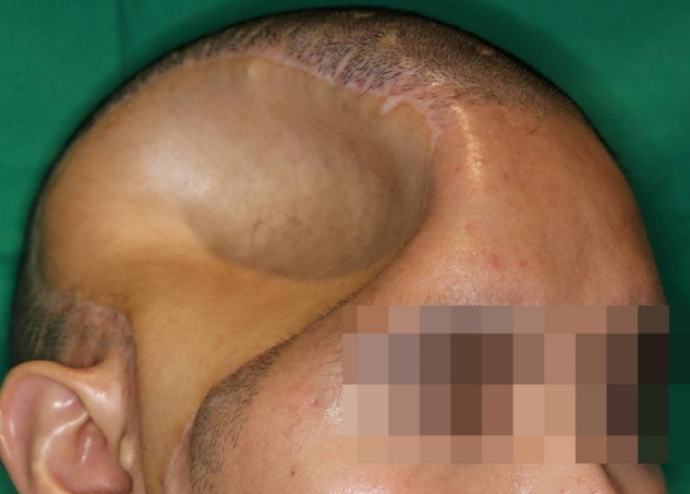
Reconstructions of extensive composite scalp and cranial defects are challenging due to high incidence of postoperative infection and reconstruction failure. In such cases, cranial reconstruction and vascularized soft tissue coverage are required. However, optimal reconstruction timing and material for cranioplasty are not yet determined. Herein, we present a large skull defect with a chronically infected wound that was not improved by repeated debridement and antibiotic treatment for 3 months. It was successfully treated with anterolateral thigh (ALT) free flap transfer for wound salvage and delayed cranioplasty with a patient-specific polyetheretherketone implant. To reduce infection risk, we performed the cranioplasty 1 year after the infection had resolved. In the meantime, depression of ALT flap at the skull defect site was observed, and the midline shift to the contralateral side was reported in a brain computed tomography (CT) scan, but no evidence of neurologic deterioration was found. After the surgery, sufficient cerebral expansion without noticeable dead-space was confirmed in a follow-up CT scan, and there was no complication over the 1-year follow-up period.
Keywords: Cranioplasty; Free tissue flap; Polyetheretherketone.
Conflict of interest statement
No potential conflict of interest relevant to this article was reported.
Figures


References
-
- Dujovny M, Fernandez P, Alperin N, Betz W, Misra M, Mafee M. Post-cranioplasty cerebrospinal fluid hydrodynamic changes: magnetic resonance imaging quantitative analysis. Neurol Res. 1997;19:311–6. - PubMed
-
- Lee JC, Kleiber GM, Pelletier AT, Reid RR, Gottlieb LJ. Autologous immediate cranioplasty with vascularized bone in high-risk composite cranial defects. Plast Reconstr Surg. 2013;132:967–75. - PubMed
-
- Reddy S, Khalifian S, Flores JM, Bellamy J, Manson PN, Rodriguez ED, et al. Clinical outcomes in cranioplasty: risk factors and choice of reconstructive material. Plast Reconstr Surg. 2014;133:864–73. - PubMed
Publication types
Grants and funding
LinkOut - more resources
Full Text Sources
