Review
. 2020 Nov 3;17(1):328.
doi: 10.1186/s12974-020-02005-x.

Cellular infiltration in traumatic brain injury

Affiliations
Review

Cellular infiltration in traumatic brain injury

Aftab Alam et al. J Neuroinflammation. .

Abstract

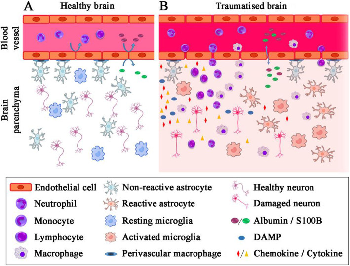
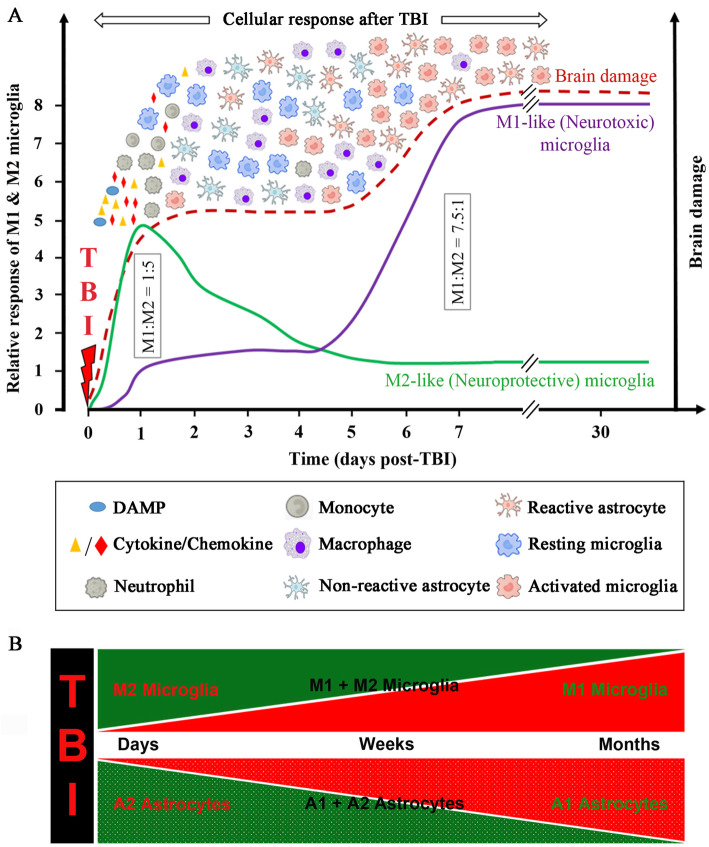
Traumatic brain injury leads to cellular damage which in turn results in the rapid release of damage-associated molecular patterns (DAMPs) that prompt resident cells to release cytokines and chemokines. These in turn rapidly recruit neutrophils, which assist in limiting the spread of injury and removing cellular debris. Microglia continuously survey the CNS (central nervous system) compartment and identify structural abnormalities in neurons contributing to the response. After some days, when neutrophil numbers start to decline, activated microglia and astrocytes assemble at the injury site-segregating injured tissue from healthy tissue and facilitating restorative processes. Monocytes infiltrate the injury site to produce chemokines that recruit astrocytes which successively extend their processes towards monocytes during the recovery phase. In this fashion, monocytes infiltration serves to help repair the injured brain. Neurons and astrocytes also moderate brain inflammation via downregulation of cytotoxic inflammation. Depending on the severity of the brain injury, T and B cells can also be recruited to the brain pathology sites at later time points.

Keywords: Cellular infiltration; Microglial dynamics; Neuroinflammation; Traumatic brain injury.


Conflict of interest statement

The authors declare that they have no competing interests.

Figures

Fig. 1
Fig. 1
Timeline of cellular response after TBI. a Upon TBI, cellular damage results in the rapid release of damage-associated molecular patterns (DAMPs) that prompt resident cells to release cytokines and chemokines. These signals quickly recruit neutrophils, which aid in the containment of the injury site and promote the removal of debris and damaged cells. As neutrophil numbers begin to decline after days, infiltrating monocytes and activated microglia and astrocytes begin to accumulate around the site of injury to perform reparative functions. Depending on the severity of the brain injury, T and B cells can also be recruited to sites of brain pathology at later time points in the response (5-7 days post-injury). Severity of the brain damage (red dashed) depends on the secondary injury mainly caused by the fluctuating microglia especially the M1-type (neurotoxic) microglia as shown by green dashed lines. b Schematic representation to highlight the behaviour of cells after TBI
Fig. 2
Fig. 2
Cellular infiltration after TBI. a In healthy brain, the functional unit constituted by firmly coupled endothelial cells and astrocytic end-feet frame the blood-brain barrier (BBB). Immune cells flow unreservedly in the blood vessel, and in the brain parenchyma, blood-borne and brain-borne proteins cannot pass into the other compartment, resting microglia survey the intact brain ecosystem. b Following TBI, the BBB disrupted/leaked activating the endothelial cells. The tight junctions between endothelial cells perish. This allows immune cells to adhere at the blood vessels lamina and then transmigration to the brain parenchyma. Specific brain proteins (e.g. S100B) are released into the blood according to their concentration gradient in exchange, serum protein enters the brain parenchyma which has been demonstrated in the cartoon. Now, microglia switch from their resting state to an activated state, embracing a phagocytic phenotype and secreting pro-inflammatory proteins. Upregulation of adhesion molecules in cerebral vessels and production of chemokines by activated microglia and astrocytes finally cause blood leukocytes to migrate into the brain parenchyma where monocytes are suggested to cause additional damage to the brain
Fig. 3
Fig. 3
Graphical representation of the cellular response and brain damage post-TBI, including the proposed microglia (M1 and M2) and astrocytes (A1 and A2) dynamics over time. a Injury to the brain may cause cell membrane disruption, vascular rupture and BBB damage. This leads to the release of DAMPs, cytokines, chemokines immediately after injury and peaks within minutes to hours and continue to release by the damaged tissue and infiltrating cells. This causes the activation of microglia and astrocytes and recruitment of circulating immune cells. These immune responses are largely overlapped (Fig. 1). The inflammatory response is the key to debris clearance, repair and regeneration post-TBI. But skewed inflammation might lead to secondary brain injury. The role of microglial activation is increasingly recognised as both a critical pathological mechanism and therapeutic target. Specifically, there is a shift from a dominance of M2 (neuroprotective) microglia into a preponderance of M1 (neurotoxic) microglia [141]. M1:M2 microglia population ratio shifts from 1:5 (day 1) to 7.5:1 (day 7) following injury [142]. Also, Wang et al. [143] whilst working with CCI mice model discovered that the phenotypic ratio of M1 and M2 at 48 h (day 2) is 1:3 and the M2-like microglia/macrophages peaked at day 5 but decreased rapidly thereafter. b Both microglia and astrocytes are highly sensitive to their environment. Unlike other inflammatory cells, astrocytes and microglia are in a constant dynamic mode. Their subtypes will shift with time and space, and with other unknown variables. Microglia and astrocytes so far have been shown to exist in two distinct reactive states (Microglia, M1-neurotoxic and M2-neuroprotective; Astrocytes, A1-neurotoxic and A2-neuroprotective). Considering these two states, it is possible that they exist as a continuum, with a heterogeneous mixed population in the middle. It should be noted that this classification is somewhat limited because microglia/macrophages can exhibit more than two canonical polarisation states [144]. In much a similar way, reactive astrocytes might have more than two polarised states. The heterogeneity of reactive microglia and astrocytes need to be investigated thoroughly

Similar articles

Cited by

References

    1. Finnie JW, Blumbergs PC. Traumatic brain injury. Vet Pathol. 2002;39:679–89. - PubMed
    1. Masel BE, DeWitt DS. Traumatic brain injury: a disease process, not an event. J Neurotrauma. 2010;27:1529–1540. doi: 10.1089/neu.2010.1358. - DOI - PubMed
    1. Davis AE. Mechanisms of traumatic brain injury: biomechanical, structural and cellular considerations. Crit Care Nurs Q. 2000;23:1–13. doi: 10.1097/00002727-200011000-00002. - DOI - PubMed
    1. Smith JS, Scholtz JM. Guanidine hydrochloride unfolding of peptide helices: separation of denaturant and salt effects. Biochemistry. 1996;35:7292–7297. doi: 10.1021/bi960341i. - DOI - PubMed
    1. Hall ED, Gibson TR, Pavel KM. Lack of a gender difference in post-traumatic neurodegeneration in the mouse controlled cortical impact injury model. J Neurotrauma. 2005;22:669–679. doi: 10.1089/neu.2005.22.669. - DOI - PubMed