. 2021 Jan 13;95(3):e01828-20.
doi: 10.1128/JVI.01828-20. Print 2021 Jan 13.

Changes in SARS-CoV-2 Spike versus Nucleoprotein Antibody Responses Impact the Estimates of Infections in Population-Based Seroprevalence Studies

Affiliations

Changes in SARS-CoV-2 Spike versus Nucleoprotein Antibody Responses Impact the Estimates of Infections in Population-Based Seroprevalence Studies

Craig Fenwick et al. J Virol. .

Abstract

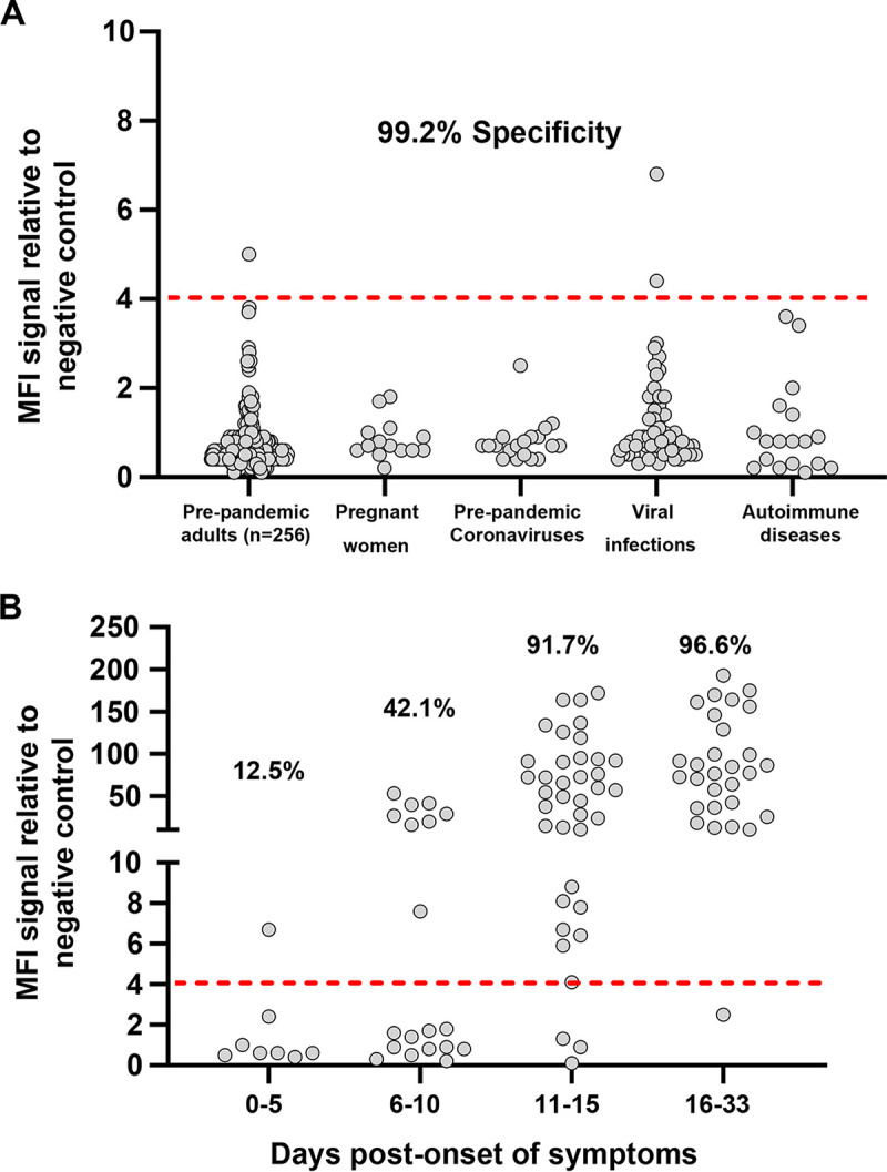
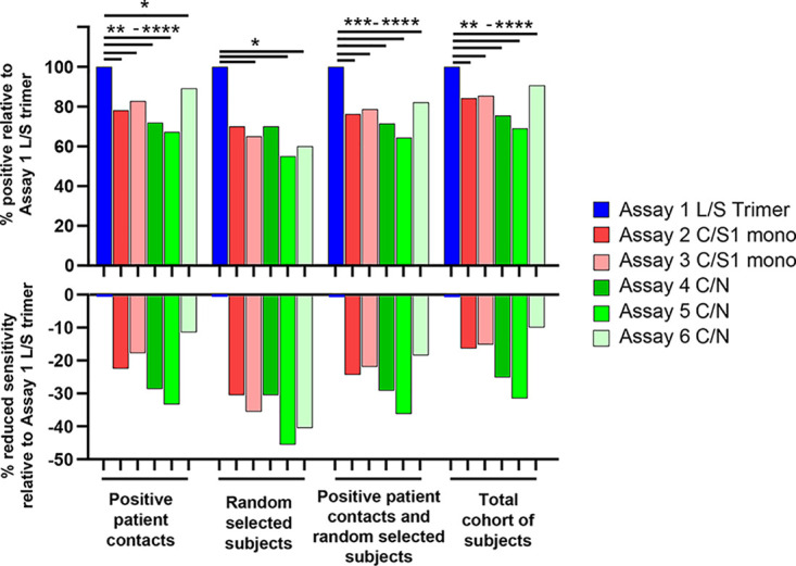
Severe acute respiratory syndrome coronavirus 2 (SARS-CoV-2)-specific antibody responses to the spike (S) protein monomer, S protein native trimeric form, or the nucleocapsid (N) proteins were evaluated in cohorts of individuals with acute infection (n = 93) and in individuals enrolled in a postinfection seroprevalence population study (n = 578) in Switzerland. Commercial assays specific for the S1 monomer, for the N protein, or within a newly developed Luminex assay using the S protein trimer were found to be equally sensitive in antibody detection in the acute-infection-phase samples. Interestingly, compared to anti-S antibody responses, those against the N protein appear to wane in the postinfection cohort. Seroprevalence in a "positive patient contacts" group (n = 177) was underestimated by N protein assays by 10.9 to 32.2%, while the "randomly selected" general population group (n = 311) was reduced by up to 45% relative to the S protein assays. The overall reduction in seroprevalence targeting only anti-N antibodies for the total cohort ranged from 9.4 to 31%. Of note, the use of the S protein in its native trimer form was significantly more sensitive compared to monomeric S proteins. These results indicate that the assessment of anti-S IgG antibody responses against the native trimeric S protein should be implemented to estimate SARS-CoV-2 infections in population-based seroprevalence studies.IMPORTANCE In the present study, we have determined SARS-CoV-2-specific antibody responses in sera of acute and postinfection phase subjects. Our results indicate that antibody responses against viral S and N proteins were equally sensitive in the acute phase of infection, but that responses against N appear to wane in the postinfection phase where those against the S protein persist over time. The most sensitive serological assay in both acute and postinfection phases used the native S protein trimer as the binding antigen, which has significantly greater conformational epitopes for antibody binding compared to the S1 monomer protein used in other assays. We believe these results are extremely important in order to generate correct estimates of SARS-CoV-2 infections in the general population. Furthermore, the assessment of antibody responses against the trimeric S protein will be critical to evaluate the durability of the antibody response and for the characterization of a vaccine-induced antibody response.

Keywords: S protein trimer; SARS-CoV-2; serology.


Figures

FIG 1
FIG 1
SARS-CoV-2-specific IgG binding antibody responses against the native trimeric S protein in a Luminex binding assay. Luminex beads covalently coupled with SARS-CoV-2 S protein trimer were used to monitor IgG-binding antibody responses in pre-COVID-19 pandemic negative-control sera and sera from SARS-CoV-2 PCR-positive donors. MFI signals for serum antibody binding were expressed as ratios compared to a negative-control pool of pre-COVID-19 pandemic human serum tested in parallel. (A) Assay specificity was evaluated using the sera from pre-COVID-19 pandemic healthy adults (n = 256; ages ranging between 18 to 81 years of age), pregnant woman (n = 14), prepandemic coronavirus-infected donors (OC43, E229, NL63, and HKU1; n = 19), patients with infectious diseases (HIV, rubella, HSV1, HSV2, RSV, CMV, EBV, influenza, and varicella; n = 57), and patients with autoimmune diseases, including lupus (n = 18). (B) The sensitivity of the S protein trimer was evaluated with sera from acute-infected SARS-CoV-2 PCR-positive donors at 0 to 5 days, 6 to 10 days, 11 to 15 days, and 16 to 33 days post-onset of symptoms. The red dashed line in A and B corresponds to the 4.0 cutoff for positivity in the IgG Luminex assay that was established by using mean value + (4 × SD) of all 364 pre-COVID-19 pandemic serum samples shown in A.
FIG 2
FIG 2
SARS-CoV-2-specific IgA-binding antibody responses against the native trimeric S protein in a Luminex binding assay. The native trimeric S protein was used to monitor IgA-binding antibodies in sera from pre-COVID-19 pandemic negative-control donors and sera from acute SARS-CoV-2 PCR-positive donors. The Luminex assay exhibited high specificity of 98.5% against a cohort of 256 prepandemic adults (left side of graph) and was effective at detecting IgA antibodies specific for S protein in most subjects at both the early stage (0 to 10 days) and later stage (11 to 33 days) post-onset of symptoms in acute PCR-positive patients (right side of graph). The red dashed line corresponds to a 6.5-fold MFI signal over the internal negative control and was established by using the mean value + (4 × SD) of the 256 pre-COVID-19 pandemic adult serum samples.
FIG 3
FIG 3
Comparative analysis of SARS-CoC-2-specific IgG binding antibody responses against S and N proteins in sera from patients with acute infection using six different serological assays. Sensitivity in detecting anti-SARS-CoV-2-specific IgG antibodies was assessed using the Luminex assay and five other commercial assays as described in the Materials and Methods section. (A) Serum samples were grouped by the number of days post initial onset of symptoms with sensitivity increasing over time. (B) Comparison in sensitivity between the different assays in samples collected from day 16 to 33 post-symptoms. L/S trimer, Luminex assay with S trimer antigen; C/S1 mono, commercial assay with S1 monomer antigen; C/N, commercial assay with nucleocapsid protein antigen.
FIG 4
FIG 4
Comparative analysis of IgG antibody responses against the trimeric S protein versus monomeric S and/or N proteins. Signal intensities for the different subject sera in the postinfection cohort were compared between the Luminex assay and the five other serological assays. Collected sera were from patients with a documented positive SARS-CoV-2 RT-PCR (90 sera; blue dots), positive patient contacts with a SARS-CoV-2 RT-PCR positive patient (177 sera; red dots), and randomly selected subjects from the general population (311 sera; green dots). Pearson correlation R2 values are given for all 578 participants (black text) or for the 183 Luminex positive sera (blue text). Regions between the dotted lines in individual graphs are indeterminate limits of response in each of the assays and above the upper limit dotted line corresponds to seropositive samples.
FIG 5
FIG 5
SARS-CoV-2-specific antibody responses to the trimeric S protein have significantly increased sensitivity compared to the S1 monomeric and/or N proteins in the postinfection population-based study. Analysis shows the percentage of seropositive subjects relative to the estimates obtained with the trimeric S protein (top) and the percentage of reduced sensitivity relative to the S1 monomeric and/or N proteins (bottom). Assays with blue bars used the S protein trimer as their bait for binding serum antibodies, while the red bars used the monomeric S1 protein and the green bars the N protein. Statistical analysis was performed using the McNemar test for matched participant samples: *, P < 0.045; **, P < 0.0022; ***, P < 0.0009; ****, P ≤ 0.0001.

References

    1. World Health Organization. 2020. Coronavirus disease (COVID-2019) situation reports-175. World Health Organization, Geneva, Switzerland.
    1. Norheim OF. 2020. Protecting the population with immune individuals. Nat Med 26:823–824. doi: 10.1038/s41591-020-0896-2. - DOI - PubMed
    1. Phelan AL. 2020. COVID-19 immunity passports and vaccination certificates: scientific, equitable, and legal challenges. Lancet 395:1595–1598. doi: 10.1016/S0140-6736(20)31034-5. - DOI - PMC - PubMed
    1. Weitz JS, Beckett SJ, Coenen AR, Demory D, Dominguez-Mirazo M, Dushoff J, Leung C-Y, Li G, Măgălie A, Park SW, Rodriguez-Gonzalez R, Shivam S, Zhao CY. 2020. Modeling shield immunity to reduce COVID-19 epidemic spread. Nat Med 26:849–854. doi: 10.1038/s41591-020-0895-3. - DOI - PMC - PubMed
    1. Okba NMA, Muller MA, Li W, Wang C, GeurtsvanKessel CH, Corman VM, Lamers MM, Sikkema RS, de Bruin E, Chandler FD, Yazdanpanah Y, Le Hingrat Q, Descamps D, Houhou-Fidouh N, Reusken C, Bosch BJ, Drosten C, Koopmans MPG, Haagmans BL. 2020. Severe acute respiratory syndrome coronavirus 2-specific antibody responses in coronavirus disease patients. Emerg Infect Dis 26:1478–1488. doi: 10.3201/eid2607.200841. - DOI - PMC - PubMed

MeSH terms