Review
. 2021 Jan-Jun:296:100017.
doi: 10.1074/jbc.REV120.013309. Epub 2020 Nov 22.

Adaptation of influenza viruses to human airway receptors

Affiliations
Review

Adaptation of influenza viruses to human airway receptors

Andrew J Thompson et al. J Biol Chem. 2021 Jan-Jun.

Abstract

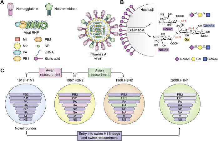
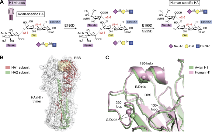
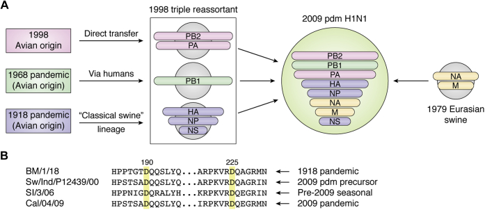
Through annual epidemics and global pandemics, influenza A viruses (IAVs) remain a significant threat to human health as the leading cause of severe respiratory disease. Within the last century, four global pandemics have resulted from the introduction of novel IAVs into humans, with components of each originating from avian viruses. IAVs infect many avian species wherein they maintain a diverse natural reservoir, posing a risk to humans through the occasional emergence of novel strains with enhanced zoonotic potential. One natural barrier for transmission of avian IAVs into humans is the specificity of the receptor-binding protein, hemagglutinin (HA), which recognizes sialic-acid-containing glycans on host cells. HAs from human IAVs exhibit "human-type" receptor specificity, binding exclusively to glycans on cells lining the human airway where terminal sialic acids are attached in the α2-6 configuration (NeuAcα2-6Gal). In contrast, HAs from avian viruses exhibit specificity for "avian-type" α2-3-linked (NeuAcα2-3Gal) receptors and thus require adaptive mutations to bind human-type receptors. Since all human IAV pandemics can be traced to avian origins, there remains ever-present concern over emerging IAVs with human-adaptive potential that might lead to the next pandemic. This concern has been brought into focus through emergence of SARS-CoV-2, aligning both scientific and public attention to the threat of novel respiratory viruses from animal sources. In this review, we summarize receptor-binding adaptations underlying the emergence of all prior IAV pandemics in humans, maintenance and evolution of human-type receptor specificity in subsequent seasonal IAVs, and potential for future human-type receptor adaptation in novel avian HAs.

Keywords: cell surface receptor; glycoprotein; hemagglutinin; influenza virus; neuraminidase; pandemic; sialic acid.


Conflict of interest statement

Conflict of interest The authors declare that they have no conflicts of interest with the contents of this article.

Figures

Figure 1
Figure 1
Cartoon schematic of influenza A virus (IAV) virion structure, receptor binding, and gene reassortment.A, major viral proteins are shown labeled, including viral surface hemagglutinin, which mediates attachment to host cell sialic acid ligands. B, human and avian receptors differ by their receptor structures/chemistries featuring predominantly α2-6 and α2-3 linkages, respectively, in the upper airway. Structures shown illustrate that sialic acid can be linked to α2-3 or α2-6 to Galβ1-4GlcNAc representing common terminal fragments of glycans that occur in nature. C, schematic of IAV reassortments leading to novel pandemic viruses during the 20th and 21st centuries. 1918 H1N1 replaced all previously circulating human IAV strains and contributed significantly to all subsequent pandemics through reassortment with avian viruses. 1918 H1N1 also entered swine species, forming a distinct swine H1 lineage that eventually contributed genes to the novel 2009 H1N1 pandemic virus.
Figure 2
Figure 2
Humanizing H1 amino acid variants and IAV hemagglutinin and receptor-binding site structures.A, effects of individual receptor-binding variants on viral receptor specificity that contributed to H1N1 entry into humans. B, overall structure of the HA (H1) hemagglutinin trimer shown as a transparent surface. HA1 and HA2 subunits from one protomer of the trimer are highlighted in red and green, respectively. C, architecture and key structural features of avian (green) and human (pink) H1. The locations of key amino acid positions are highlighted through depiction of Cα positions as enlarged spheres and via labeling of key residues and secondary structural features. Assembled in Pymol (Schrodinger LLC) using PDB IDs: 1RV0 and 1RVZ.
Figure 3
Figure 3
Reassortment leading to emergence of the 2009 pandemic H1N1 virus.A, cartoon schematic of various swine lineages contributing gene segments to the novel virus (adapted from data in Garten et al. (2009) (24)). B, conservation of key humanizing receptor-binding variants in example viruses from the original 1918 pandemic, through the classical swine H1N1 lineage, and within both pre- and post-2009 human H1N1 strains. Strain abbreviations are: A/Brevig Mission/1/1918, A/swine/Indiana/P12439/2000 (nearest identified swine HA gene precursor to 2009 human pandemic strains), A/Solomon Islands/3/2006 (pre-2009 seasonal), and A/California/04/2009 (2009 pandemic).
Figure 4
Figure 4
Human-adaptive receptor-binding variants underlying the 1957 H2N2 and 1968 H3N2 pandemic viruses.A, similar to 1918 H1N1, H2 and H3 HAs feature a single dominant variant (Q226L) enabling binding to human-type receptors and a stabilizing covariant (G228S) that ensures specificity. B, architecture and key structural features of human H1 (pink) and human H2 (lilac). The locations of key amino acid positions are highlighted through depiction of Cα positions as enlarged spheres and via labeling of key residues and secondary structural features. The key 226 and 228 positions in H2/H3 lie in close proximity to D225 in human H1. C, enlarged overlay of the avian (yellow) and human (gray) H3 RBS. Panels (B) and (C) assembled in Pymol (Schrodinger LLC) using PDB IDs: 1RVZ, 3KU6, 1MQM, & 6TZB.
Figure 5
Figure 5
Length-selective binding of contemporary human H3N2 viruses. Glycan microarray segments are shown comparing binding of A/Hong Kong/1/68 (novel H3N2 pandemic strain) with A/Victoria/361/2011 (adapted human seasonal strain) to exclusively α2-6-linked N-glycans that vary only by length through the number of N-acetyllactosamine (LacNAc) repeats beneath the terminal sialic acid. Vic/11 binds exclusively to N-glycans featuring at least 3 LacNAc units. Note: for compounds 113 to 119, biantennary N-glycans, (LacNAc)2 and (LacNAc)3 structures are duplicated (114 & 115 and 116 & 117, respectively) with slightly different chemical attachment linkers. Figure adapted from data published in Peng et al. (2017) (49) with permission from Cell Press, see manuscript for further details.
Figure 6
Figure 6
Receptor-binding adaptations are dictated by the conformation of receptor itself.A, cartoon and isolated receptor structures showing respective low-energy linear and “hook”-shaped conformations of α2-3 and α2-6 sialosides, respectively. CD, human-adaptive variants favor accommodation of the α2-6-linked receptor conformation and lead to hindrance of linear α2-3 receptors. C, the smaller D190 side chain increases stability and reduces clashing with human-type receptors, while D225 adds a new H-bonding interaction. D, Q226 favors binding of galactose in the linear avian-type conformation through H-bonding interactions. E, mutation of Q226 to leucine (L) removes favorable α2-3-binding H-bonds while the new hydrophobic side chain hinders sugars lying directly over this position. Panel (A) assembled using CCP4Mg (148), panels (CD) assembled in Pymol (Schrodinger LLC) using PDB IDs: 1RVZ, 1MQM, & 6TZB.

References

    1. Krammer F., Smith G.J.D., Fouchier R.A.M., Peiris M., Kedzierska K., Doherty P.C., Palese P., Shaw M.L., Treanor J., Webster R.G., Garcia-Sastre A. Influenza. Nat. Rev. Dis. Primers. 2018;4:3. - PMC - PubMed
    1. Taubenberger J.K., Kash J.C., Morens D.M. The 1918 influenza pandemic: 100 years of questions answered and unanswered. Sci. Transl. Med. 2019;11 - PMC - PubMed
    1. Thompson A.J., de Vries R.P., Paulson J.C. Virus recognition of glycan receptors. Curr. Opin. Virol. 2019;34:117–129. - PMC - PubMed
    1. Shinya K., Ebina M., Yamada S., Ono M., Kasai N., Kawaoka Y. Avian flu: influenza virus receptors in the human airway. Nature. 2006;440:435–436. - PubMed
    1. Rogers G.N., Paulson J.C. Receptor determinants of human and animal influenza virus isolates: differences in receptor specificity of the H3 hemagglutinin based on species of origin. Virology. 1983;127:361–373. - PubMed

Publication types

MeSH terms