

Giant Intrathoracic Schwannoma: A Case Report
- PMID: 33144463
- PMCID: PMC7811636
- DOI: 10.21873/invivo.12194
Giant Intrathoracic Schwannoma: A Case Report
Abstract
Background/aim: Thoracic neurogenic tumors are most frequently located in the posterior part of the mediastinum or on the chest wall, along the intercostal nerves. Schwannomas are very well tolerated for a long period, until the tumor reaches a large size and compression of the neighbouring mediastinal organs, chest wall or spine appears. The purpose of this article was to present a case of a giant right forth intercostal nerve Schwannoma, completely resected by a right antero-lateral thoracotomy. In addition, intrathoracic giant neurogenic tumors are a rarity.
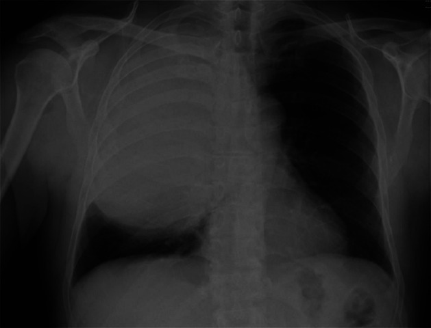
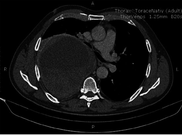
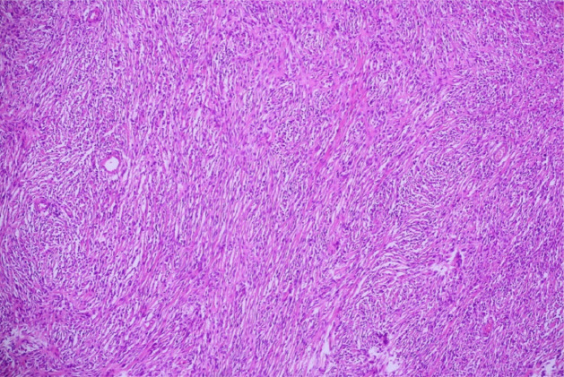
Case report: The patient presented with only diminished tolerance to physical activity with no other obvious symptoms. Standard chest radiography revealed a well-defined opacity of subcostal intensity, occupying two thirds of the right hemithorax, forming a common body with the mediastinal shadow. Thoracic computed tomography (CT) identified a 21/11 cm solid mass that compresses the right lung and the right main bronchus with both a solid component and a central liquid area. Open surgery was performed in order to remove the tumor, which was 20.5/12.5/9 cm in size and weighed 1,830 g, well defined, with no invasion of the adjacent organs, having a solid-fibromatous aspect as well as a central necrotic area. The origin of the tumor was confirmed from the posterolateral part of the forth intercostal nerve. Pathology examination and immunohistochemistry confirmed the diagnosis of a benign Schwannoma.
Conclusion: Benign intrathoracic Schwannomas are asymptomatic for long periods and the main therapeutic option is complete surgical resection. The surgical approach, either open or video-assisted is dictated by the localisation of the tumor, local extension and most importantly the size of the neurogenic mass.
Keywords: Schwannoma; anterolateral thoracotomy; dumbbell tumor; giant neurogenic tumors.
Copyright© 2020, International Institute of Anticancer Research (Dr. George J. Delinasios), All rights reserved.
Conflict of interest statement
The Authors declare no conflicts of interest regarding this study.
Figures
References
-
- Yamaguchi M, Yoshino I, Fukuyama S, Osoegawa A, Kameyama T, Tagawa T, Maehara Y. Surgical treatment of neurogenic tumors of the chest. Ann Thorac Cardiovasc Surg. 2004;10(3):148–151. - PubMed
Publication types
MeSH terms
LinkOut - more resources
Full Text Sources