. 2020 Nov 3;11(1):5547.
doi: 10.1038/s41467-020-19325-4.

Nearest-neighbor NMR spectroscopy: categorizing spectral peaks by their adjacent nuclei

Affiliations

Nearest-neighbor NMR spectroscopy: categorizing spectral peaks by their adjacent nuclei

Soumya P Behera et al. Nat Commun. .

Abstract

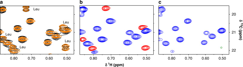
Methyl-NMR enables atomic-resolution studies of structure and dynamics of large proteins in solution. However, resonance assignment remains challenging. The problem is to combine existing structural informational with sparse distance restraints and search for the most compatible assignment among the permutations. Prior classification of peaks as either from isoleucine, leucine, or valine reduces the search space by many orders of magnitude. However, this is hindered by overlapped leucine and valine frequencies. In contrast, the nearest-neighbor nuclei, coupled to the methyl carbons, resonate in distinct frequency bands. Here, we develop a framework to imprint additional information about passively coupled resonances onto the observed peaks. This depends on simultaneously orchestrating closely spaced bands of resonances along different magnetization trajectories, using principles from control theory. For methyl-NMR, the method is implemented as a modification to the standard fingerprint spectrum (the 2D-HMQC). The amino acid type is immediately apparent in the fingerprint spectrum. There is no additional relaxation loss or an increase in experimental time. The method is validated on biologically relevant proteins. The idea of generating new spectral information using passive, adjacent resonances is applicable to other contexts in NMR spectroscopy.


Conflict of interest statement

The authors declare no competing interests.

Figures

Fig. 1
Fig. 1. Theory of decoupling using selective inversion to distinguish leucine from valine.
a Trajectory of the 13CH3 leucine (red) and valine (blue, dashed) magnetization during a constant time encoding period of duration T = 1/1JCC = 28 ms. Selective decoupling of valine at time T/2 refocuses the coupling and produces positive peaks for valine at time T. Leucine is not decoupled, and the coupling interaction leads to negative peaks at time T. b The distributions of Cβ and Cγ chemical shifts of valine (blue) and leucine (red) are distinct. Assuming an initial state of Iz, our decoupling pulse selectively inverts the CβVal, which selectively decouples the valine methyl resonance. The encoding of the methyl-carbon chemical shift continues unaffected during the selective homonuclear decoupling pulse.
Fig. 2
Fig. 2. Testing the selective decoupling pulse using real time and constant-time 1H–13C HMQC on MBP.
a Overlay of a section of a real-time 2D 1H–13C methyl HMQC experiment with (orange) and without (black) the selective decoupling pulse on a 500 µM MBP (42.5 kDa) protein. The leucine peaks remain as doublets, whereas the valine peaks collapse into singlets. b Constant-time 1H–13C HMQC experiment with indirect constant-time period set to T = 28 ms (1/1JCC) and shaped pulse applied at time T/2. Valine resonances are positive (blue) whereas leucine peaks are negative (red). c A version of the experiment with constant-time evolution period is set to T = 42 ms (3/(2 1JCC)) completely eliminates the leucine peaks from the spectrum with positive valine peaks remaining (blue). Full spectra are in Supplementary Fig. 5.
Fig. 3
Fig. 3. Schematic explaining disappearance of isoleucine and leucine peaks when the constant-time duration is set to T = 3/(2 1JCC).
The simulation shows the transverse projection of the 13CH3 Leu (red), Val (blue) magnetization vector, with arrows showing the sense of rotation with respect to the intrinsic chemical shift frequency. The resulting 13CH3-x magnetization for Leu (red), Val (blue, dashed) are shown below. The constant-time evolution period T is set to 42 ms (3/(2 1JCC)). The shaped pulse at the T/2 selectively decouples CγVal from CβVal, resulting in the appearance of the valine peaks. The disappearance of isoleucine and leucine peaks in the spectrum is due to the cancelling out of the anti-phase term arising because of the 13C–13C spin coupling.
Fig. 4
Fig. 4. Application of the valine-selective decoupling pulse on biologically relevant proteins.
Constant-time 2D 1H–13C methyl HMQC spectra of four biologically relevant proteins are shown. a Cas9 HNH domain (130 µM, 15 kDa); b Cas9 REC1-2 domain (250 µM, 52 kDa); c Human IL2 (920 µM, 15.4 kDa); d eIF4A (50 µM, 46 kDa). Unambiguous distinction between the valine (blue) and leucine (red) resonances is achieved. Only Leu–Val overlapping region of the HMQC spectrum is shown here. The full spectra of these proteins are presented in Supplementary Figs. 6 and 7. e Here we show the 3D structure for the REC1-2 domain of Cas9 (PDB ID: 4UN3) where the valine and leucine residues are highlighted in blue and red, respectively.

Similar articles

Cited by

References

    1. Baldwin AJ, Kay LE. NMR spectroscopy brings invisible protein states into focus. Nat. Chem. Biol. 2009;5:808–814. doi: 10.1038/nchembio.238. - DOI - PubMed
    1. Barrett PJ, et al. The quiet renaissance of protein nuclear magnetic resonance. Biochemistry. 2013;52:1303–1320. doi: 10.1021/bi4000436. - DOI - PMC - PubMed
    1. Bax A, Clore GM. Protein NMR: boundless opportunities. J. Magn. Reson. 2019;306:187–191. doi: 10.1016/j.jmr.2019.07.037. - DOI - PMC - PubMed
    1. Kay LE. Solution NMR spectroscopy of supra-molecular systems, why bother? A methyl-TROSY view. J. Magn. Reson. 2011;210:159–170. doi: 10.1016/j.jmr.2011.03.008. - DOI - PubMed
    1. Alderson TR, Kay LE. Unveiling invisible protein states with NMR spectroscopy. Curr. Opin. Struct. Biol. 2020;60:39–49. doi: 10.1016/j.sbi.2019.10.008. - DOI - PubMed

Publication types