. 2020 Nov 3;11(1):5559.
doi: 10.1038/s41467-020-19032-0.

A trimeric Rab7 GEF controls NPC1-dependent lysosomal cholesterol export

Affiliations

A trimeric Rab7 GEF controls NPC1-dependent lysosomal cholesterol export

Dick J H van den Boomen et al. Nat Commun. .

Abstract

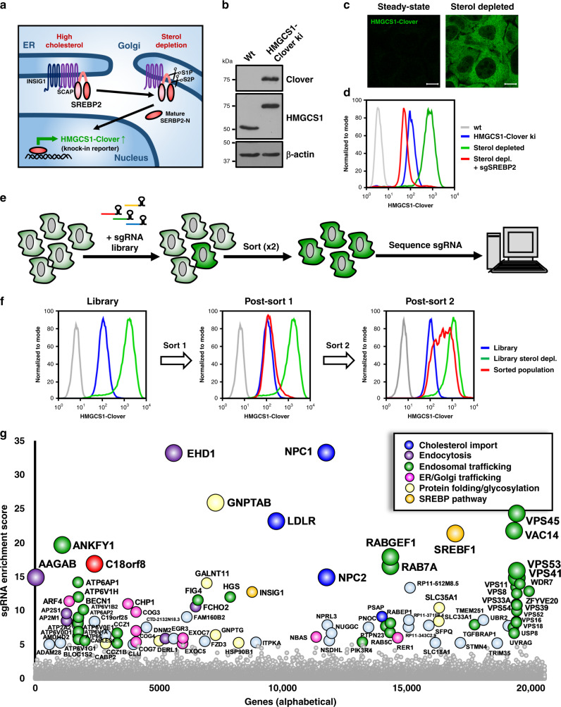
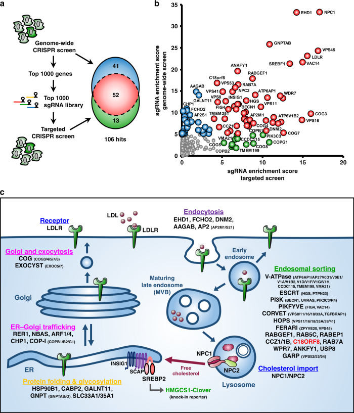
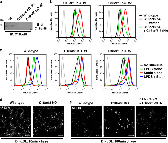
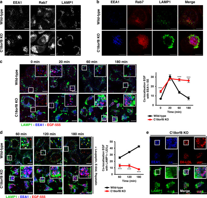
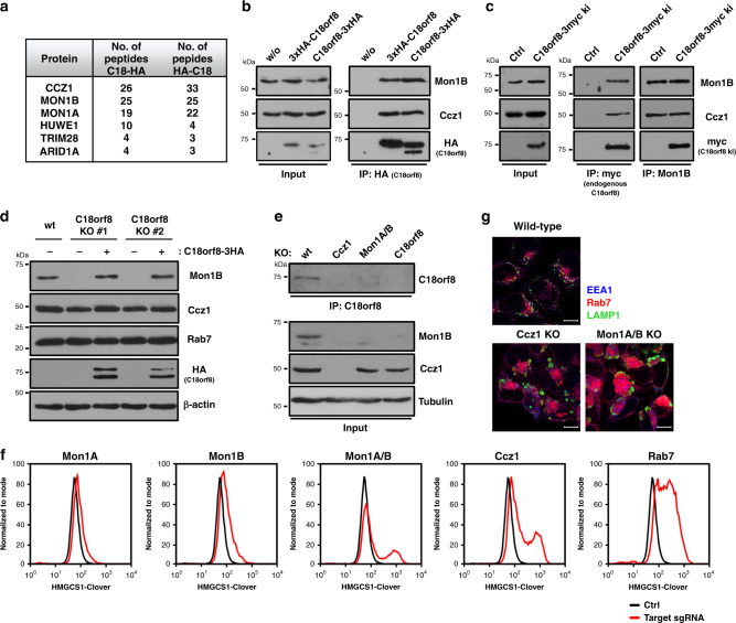
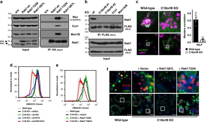
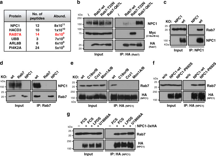
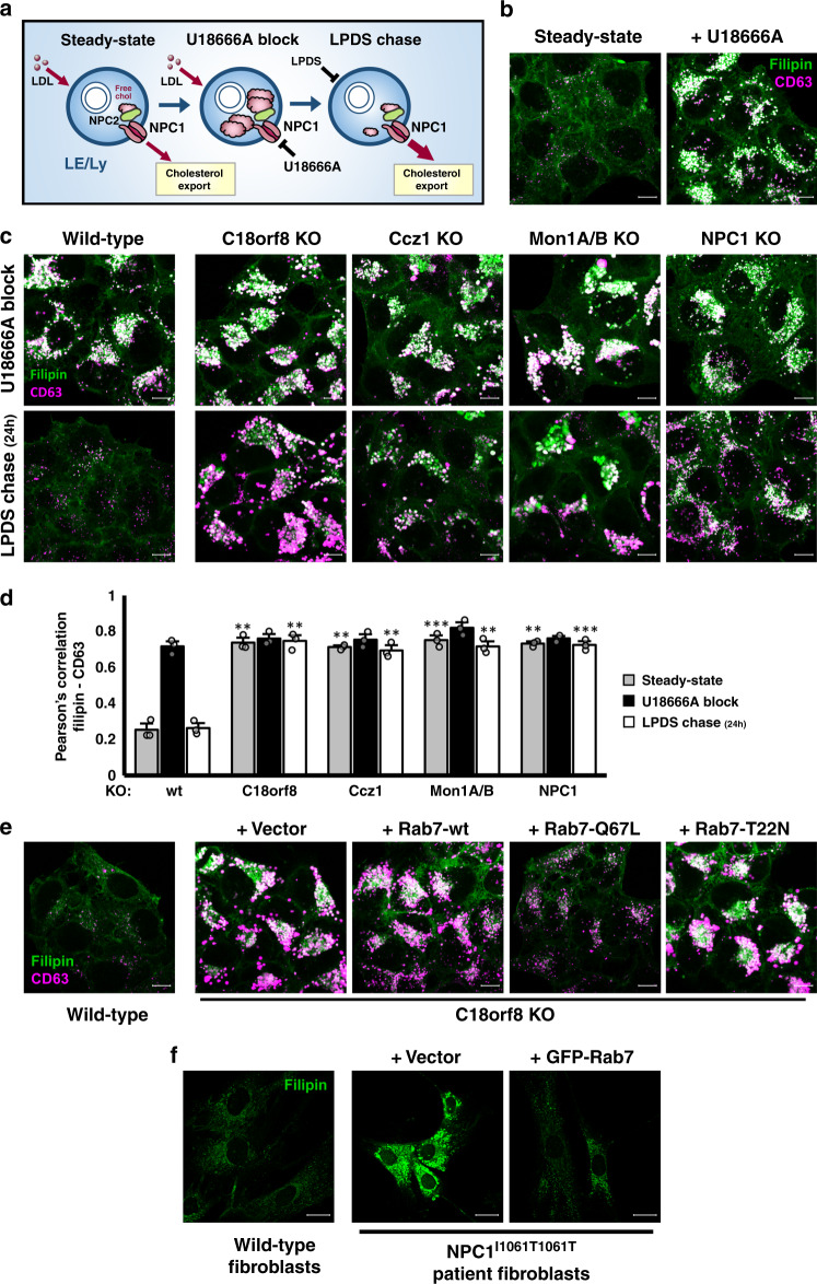
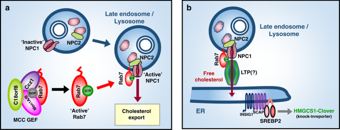
Cholesterol import in mammalian cells is mediated by the LDL receptor pathway. Here, we perform a genome-wide CRISPR screen using an endogenous cholesterol reporter and identify >100 genes involved in LDL-cholesterol import. We characterise C18orf8 as a core subunit of the mammalian Mon1-Ccz1 guanidine exchange factor (GEF) for Rab7, required for complex stability and function. C18orf8-deficient cells lack Rab7 activation and show severe defects in late endosome morphology and endosomal LDL trafficking, resulting in cellular cholesterol deficiency. Unexpectedly, free cholesterol accumulates within swollen lysosomes, suggesting a critical defect in lysosomal cholesterol export. We find that active Rab7 interacts with the NPC1 cholesterol transporter and licenses lysosomal cholesterol export. This process is abolished in C18orf8-, Ccz1- and Mon1A/B-deficient cells and restored by a constitutively active Rab7. The trimeric Mon1-Ccz1-C18orf8 (MCC) GEF therefore plays a central role in cellular cholesterol homeostasis coordinating Rab7 activation, endosomal LDL trafficking and NPC1-dependent lysosomal cholesterol export.


Conflict of interest statement

The authors declare no competing interests.

Figures

Fig. 1
Fig. 1. A genome-wide CRISPR screen identifies essential factors in cellular cholesterol homeostasis.
a, b Creation of a HMGCS1-Clover CRISPR knock-in reporter in HeLa cells. a Schematic representation of the HMGCS1-Clover knock-in reporter and its induction upon sterol depletion by the SREBP2 transcription factor. b Immunoblotting of a reporter clone shows the endogenous HMGCS1-Clover fusion protein, detected by both HMGCS1- and GFP/Clover-specific antibody staining. c, d Sterol depletion induces HMGCS1-Clover expression in an SREBP-dependent manner. c HeLa HMGCS1-Clover cells were sterol depleted overnight and HMGCS1-Clover expression was analysed by microscopy. Representative images are shown from five fields per condition. Scale bars = 10 µm. d HeLa HMGCS1-Clover CAS9 cells were transfected with sgRNA against SREBF2 (red line) or control (blue and green lines), sterol depleted at day 7 (green and red lines) and HMGCS1-Clover expression was determined by flow cytometry. e–g Genome-wide CRISPR screening using the HMGCS1-Clover cholesterol reporter. e Schematic for the identification of genes essential for cellular cholesterol homeostasis. f HeLa HMGCS1-Clover/Cas9 cells were transduced with a genome-wide sgRNA library (220,000 sgRNAs). Rare HMGCS1- Cloverhigh cells were isolated using two rounds of cell sorting, resulting in a 60% enriched HMGCS1-Cloverhigh population (right panel, red line). g Illumina sequencing of sgRNAs in the isolated HMGCS1-Cloverhigh population shows sgRNA enrichment for genes involved in cholesterol uptake (LDLR, NPC1, NPC2; blue), protein folding and glycosylation (yellow), membrane trafficking of LDLR/LDL (pink, purple, green) and SREBP2 function (orange). Genes with MAGeCK sgRNA enrichment score < 10−5 are indicated by enrichment score and gene name. The full dataset is available in Supplementary Data 1.
Fig. 2
Fig. 2. A secondary sub-genomic CRISPR screen validates genes required for cellular LDL-cholesterol uptake.
a Schematic of the primary (genome-wide) and secondary (targeted, top 1000) screening approach. Screening results from both screens show extensive overlap and create a complementary dataset of 106 genes required for cellular cholesterol homeostasis. b Comparison of the genome-wide (vertical) and targeted (horizontal) CRISPR screening results. Genes are indicated by MAGeCK sgRNA enrichment score, with hits specific to the genome-wide screen (score < 10−5) indicated in blue, hits specific to the targeted screen (score <10-4) in green and hits in both screens in red. A select number of hits is indicated by gene name. The full datasets are available in Supplementary Fig. 2 and Supplementary Data 1. c A schematic representation of combined hits from the genome-wide and targeted CRISPR screens highlights the central role of LDL-cholesterol import in cellular cholesterol homeostasis. Hits are grouped by membrane trafficking pathway of their involvement. A full list of hits from both screens is available in Supplementary Data 1.
Fig. 3
Fig. 3. C18orf8 is required for endosomal LDL-cholesterol uptake.
a, b C18orf8-deficient cells show spontaneous cholesterol deficiency. a Wild-type and C18orf8-deficient clones were lysed, endogenous C18orf8 immunoprecipitated, and detected by a C18orf8-specific antibody. b C18orf8-deficient HMGCS1-Clover clones were transduced with HA-tagged C18orf8 (C18orf8-3xHA; green lines) or empty vector (red lines) and HMGCS1-Clover expression was determined by flow cytometry at day 18 using wild-type HMGCS1-Clover cells as a control (black lines) (three independent experimental replicates). c C18orf8-deficient cells are dependent on endogenous cholesterol biosynthesis. Wild-type and C18orf8-deficient HMGCS1-Clover cells were either cultured in LPDS to block exogenous LDL-cholesterol uptake (green lines), treated with mevastatin to block endogenous cholesterol biosynthesis (red lines), or a combination of both treatments (blue lines), after which cells were analysed by flow cytometry for HMGCS1-Clover expression. d, e C18orf8-deficient cells show defective endo-lysosomal LDL degradation. Wild-type, C18orf8-deficient or C18orf8-deficient cells complemented with C18orf8-3xHA were starved for 1 h and pulse-labelled with fluorescent Dil-LDL, incubated for 5 min (d) or 180 min (e), fixed and visualised by confocal microscopy. Exposure times were kept constant between individual conditions at given time points. Representative images are shown from five fields per condition. Scale bars = 10 µm.
Fig. 4
Fig. 4. C18orf8-deficient cells show severe defects in late endosome morphology and early-to-late endosomal trafficking.
a, b C18orf8-deficient cells show clustering of EE and swelling of LE/Ly. Confocal microscopy comparing EEA1, Rab7 and LAMP1 single (a) or co-staining (b) in wild-type versus C18orf8-deficient cells. Representative images are shown from five fields per condition and 2 independent experiments. c–e C18orf8-deficient cells have severe defects in early-to-late endosomal trafficking. c, d Wild-type and C18orf8-deficient cells were starved for 1 h, pulse-labelled with AlexaFluor555-conjugated EGF (red), incubated for the indicated times, fixed and stained for EEA1 (blue) and LAMP1 (green). In d a cocktail of protease inhibitors (Leupeptin/E-64d/Pepstatin) was added during starve and chase to block EGF degradation. EEA1+ and LAMP1+ vesicles were identified using Volocity software and the percentage of EGF co-localising with these structures was determined from n = 5 fields per condition with at least eight cells per field. Error bars reflect standard error of mean (two-sided unpaired t-test, ***p < 0.001). e C18orf8-deficient cells were stimulated with Dil-LDL (red), chased for 3 h, fixed and stained for EEA1 (blue) and LAMP1 (green). Representative images are shown from five fields. Scale bars = 10 µm.
Fig. 5
Fig. 5. C18orf8 forms an integral component of the Mon1-Ccz1 (MC1) complex, essential for complex stability and function.
a–c C18orf8 interacts with the Mon1-Ccz1 complex. Immune precipitations of exogenous 3xHA-C18orf8 (N-term) or C18orf8-3xHA (C-term) were analysed by mass spectrometry (a) or Western blotting using Mon1B-, Ccz1- and HA-specific antibodies (b). Proteins detected by >3 peptides in both C18orf8 samples and absent from control, are indicated in a. Full MS results are available in Supplementary Data 2 and via ProteomeXchange with identifier PXD021444. c Immune precipitation of an endogenous C18orf8-3xMyc fusion or Mon1B reveals a reciprocal interaction between endogenous C18orf8, Ccz1 and Mon1B (see also Supplementary Fig. 5c). d, e C18orf8, Mon1B and Ccz1 show reciprocal stabilisation. Immunoblot analysis of lysates from (d) wild-type, C18orf8-deficient and C18orf8-deficient cells complemented with C18orf8-3xHA (3 independent experimental replicates); or (e) wild-type, C18orf8-, Ccz1- and Mon1A/B-deficient cells. For endogenous C18orf8 detection, C18orf8 was immune precipitated prior to immunoblotting (e). f, g Ccz1- and Mon1A/B-deficient cells show cholesterol deficiency and disruption of LE/Ly morphology. f HMGCS1-Clover CAS9 cells (black lines) were transfected with sgRNAs against Mon1A, Mon1B, Mon1A and Mon1B, Ccz1 or Rab7 (red lines) or control (black lines), grown for 14 days, treated overnight with mevastatin and analysed for HMGCS1-Clover expression. g Wild-type, Ccz1- and Mon1A/B-deficient cells were stained intracellularly for EEA1 (blue), Rab7 (red) and LAMP1 (green). Representative images are shown from five fields per condition. Scale bars = 10 µm.
Fig. 6
Fig. 6. The trimeric Ccz1-Mon1-C18orf8 (MCC) complex activates mammalian Rab7.
a The MCC complex binds an inactive Rab7 (T22N). Immune precipitations of 2xHA-tagged wild-type, T22N or Q67L Rab7 from C18orf8-3xMyc knock-in cells, were analysed by immunoblot using Myc-, Ccz1- and Mon1B-specific antibodies (2 independent experimental replicates, see also Supplementary Fig. 6a). b, c C18orf8-, Ccz1- and Mon1A/B-deficient cells lack activation-dependent recruitment of Rab7 effectors. b Immune precipitations of 3xFLAG-RILP from wild-type, C18orf8-, Ccz1- or Mon1A/B-deficient cells were analysed by immunoblotting for endogenous Rab7 (two independent experimental replicates). c Wild-type and C18orf8-deficient cells were transfected with HA-RILP or ORP1L (Supplementary Fig. 7a) and stained intracellularly for HA (green) and LAMP1 (magenta). Mander’s correlation was determined for n = 5 fields per condition from two independent experiments. Error bars reflect standard deviation (two-sided unpaired t-test, p = 2.1 × 10−5, ***p < 0.001). df Cholesterol and trafficking defects in C18orf8-deficient cells can be rescued by knockdown of Rab7GAPs or expression of a constitutively active Rab7 (Q67L). d HMGCS1-Clover expression was determined at day 8 after transduction of C18orf8-deficient cells with shRNAs against TBC1D5 (green), TBC1D15 (blue) or both (red). e-f C18orf8-deficient cells were transduced with 2xHA-tagged Rab7-T22N (green), -Q67L (red) or an empty vector (black) and either (e) analysed at day 10 by flow cytometry for HMGCS1-Clover expression (two independent experimental replicates, see also Supplementary Fig 6b), or f pulse-labelled with Dil-LDL (red), incubated for 3 h and stained intracellularly for LAMP1 (green) and EEA1 (blue). Representative images are shown from 5 fields per condition. Scale bars = 10 µm.
Fig. 7
Fig. 7. C18orf8-, Ccz1- and Mon1A/B- (MCC)-deficient cells accumulate free cholesterol in a swollen lysosomal compartment.
a Filipin staining of wild-type, C18orf8-deficient and complemented C18orf8-deficient cells; or c wild-type, Ccz1- and Mon1A/B-deficient cells. b Filipin (green) co-staining with the LE/Ly markers Rab7 (blue) and LAMP1 (red) in C18orf8-deficient cells. d Theonellamides (TNM) immuno-gold labelling of wild-type and C18orf8-deficient cells, visualised by EM. Multivesicular bodies (MVBs) are indicated by arrows. Note MVBs are markedly enlarged in C18orf8-deficient cells. e Filipin staining of wild-type, C18orf8- and NPC1-deficient cells. All confocal microscopy images are representative of five fields per condition and two independent experiments. Scale bars for confocal microscopy = 10 µm, scale bars for EM = 200 nm.
Fig. 8
Fig. 8. Rab7 interacts with the lysosomal cholesterol transporter NPC1 in an activation-dependent manner.
a Immune-precipitation of HA-tagged NPC1 and detection of NPC1-interacting proteins using mass spectrometry. Most abundant interaction partners detected with >2 peptides are indicated (full dataset available in Supplementary Data 2 and via ProteomeXchange with identifier PXD021444). b Immune precipitations of HA-tagged wild-type, dominant-negative (T22N) or constitutively active Rab7 (Q67L) show an activation-dependent interaction between Rab7 and endogenous NPC1 (two independent experimental replicates). c, d Reciprocal immune-precipitation of endogenous NPC1 (c) or Rab7 (d) shows a strong interaction between both proteins. e The Rab7-NPC1 interaction is lost in MCC-deficient cells that lack Rab7 activation. Wild-type, C18orf8-, Ccz1- and Mon1A/B-deficient cells were stably transduced with the inactive NPC1-P692S-HA. HA-tagged NPC1 was immune precipitated and immune blotted for endogenous Rab7. The inactive NPC1-P692S was used to prevent altering lysosomal cholesterol content by NPC1 overexpression. (two independent experimental replicates) f, g The Rab7-NPC1 interaction is independent of NPC1 activity or lysosomal cholesterol levels. f NPC1-deficient cells were complemented with HA-tagged wild-type or inactive P692S-mutant NPC1 and NPC1-HA immune-precipitations were analysed by immune blotting for endogenous Rab7. g Wild-type NPC1-HA complemented cells were treated with LPDS to decrease, or U18666A to increase lysosomal cholesterol levels and the NPC1-Rab7 interaction was probed using immune precipitation (two independent experimental replicates).
Fig. 9
Fig. 9. Rab7 activation by the MCC GEF controls NPC1-dependent lysosomal cholesterol export.
a–d Lysosomal cholesterol export is abolished in NPC1-, C18orf8-, Ccz1- and Mon1A/B-deficient cells. a Schematic depiction of the cholesterol export assay. Similar to a pulse-chase analysis, LDL-derived free cholesterol is allowed to accumulate in a lysosomal compartment using the NPC1 inhibitor U18666A (pulse) and released during the LPDS chase. b Free cholesterol (Filipin, green) accumulates in a CD63+ LE/Ly compartment (magenta) of wild-type HeLa cells following 24 h incubation with U18666A. c Wild-type, NPC1-, C18orf8-, Ccz1- and Mon1A/B-deficient cells were treated for 24 h with U18666A (top panels), followed by a 24 h chase in the presence of LPDS and mevastatin (lower panels). Lysosomal cholesterol accumulation was visualised using Filipin (green) co-staining with the LE/Ly marker CD63 (magenta). d Co-localisation of Filipin with CD63 is plotted as Pearson correlation, calculated from n = 3 independent experiments with six fields per condition per experiment and >8 cells per field. Error bars reflect standard error of mean (two-side paired t-test, **p < 0.01, ***p < 0.001). e Cholesterol accumulation in C18orf8-deficient cells is abolished by overexpression of a hyperactive Rab7. C18orf8-deficient cells were transduced with a wild-type, dominant-negative (T22N) or constitutively active Rab7 (Q67L) or empty vector and co-stained with Filipin (green) and anti-CD63 (magenta). Representative images are shown from five fields per condition. f Rab7 overexpression rescues lysosomal cholesterol accumulation in NPC patient fibroblasts. NPC1I1061T/I1061T primary patient fibroblasts were transduced with a GFP-tagged wild-type Rab7 or vector control and analysed at day 7 for cholesterol accumulation using Filipin staining. Fibroblasts from a healthy individual were used as a control. Representative images are shown from five fields per condition and two independent experiments (see also Supplementary Fig. 9). Scale bars = 10 µm.
Fig. 10
Fig. 10. A trimeric Rab7 GEF controls NPC1-dependent lysosomal cholesterol export.
Model for MCC and Rab7 function in lysosomal cholesterol export. The trimeric Mon1-Ccz1-C18orf8 (MCC) GEF activates mammalian Rab7, which binds the NPC1 cholesterol transporter and either a) directly activates NPC1’s cholesterol export function; or b) assembles a down-stream membrane contact site (MCS) at which a yet-uncharacterised lipid transfer protein (LTP) mediates cholesterol transfer to the ER and/or plasma membrane. A combined Rab7 function in NPC1 activation and MCS formation would assure lysosomal cholesterol export only proceeds once a down-stream lipid transfer module is assembled.

References

    1. Brown MS, Goldstein JL. The SREBP pathway: regulation of cholesterol metabolism by proteolysis of a membrane-bound transcription factor. Cell. 1997;89:331–340. doi: 10.1016/S0092-8674(00)80213-5. - DOI - PubMed
    1. Lund-Katz S, Phillips MC. Packing of cholesterol molecules in human low-density lipoprotein. Biochemistry. 1986;25:1562–1568. doi: 10.1021/bi00355a016. - DOI - PubMed
    1. Goldstein JL, Brown MS. The LDL receptor. Arterioscler Thromb. Vasc. Biol. 2009;29:431–438. doi: 10.1161/ATVBAHA.108.179564. - DOI - PMC - PubMed
    1. Pfeffer SR. NPC intracellular cholesterol transporter 1 (NPC1)-mediated cholesterol export from lysosomes. J. Biol. Chem. 2019;294:1706–1709. doi: 10.1074/jbc.TM118.004165. - DOI - PMC - PubMed
    1. Watari H, et al. Mutations in the leucine zipper motif and sterol-sensing domain inactivate the Niemann-Pick C1 glycoprotein. J. Biol. Chem. 1999;274:21861–21866. doi: 10.1074/jbc.274.31.21861. - DOI - PubMed

Publication types

MeSH terms