. 2020 Nov 3;11(11):947.
doi: 10.1038/s41419-020-03155-9.

Targeting miR-18a sensitizes chondrocytes to anticytokine therapy to prevent osteoarthritis progression

Affiliations

Targeting miR-18a sensitizes chondrocytes to anticytokine therapy to prevent osteoarthritis progression

Chengjie Lian et al. Cell Death Dis. .

Abstract

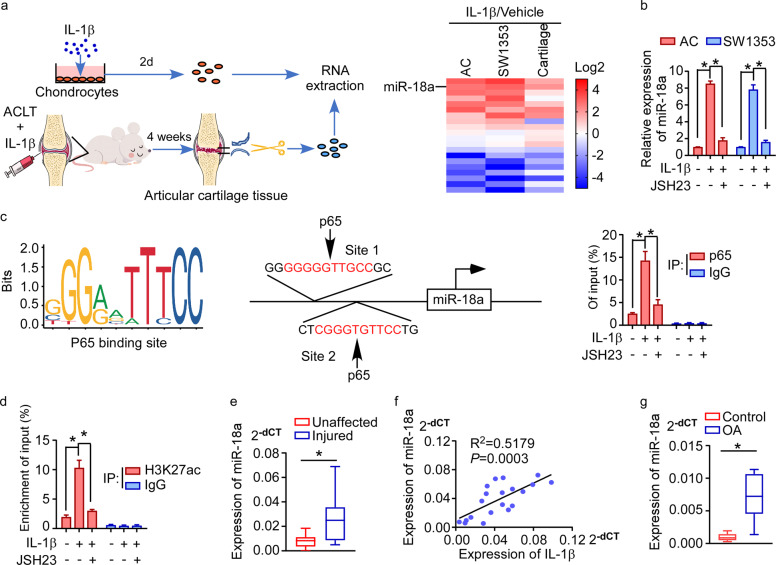
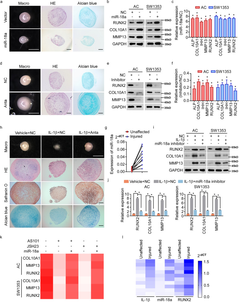
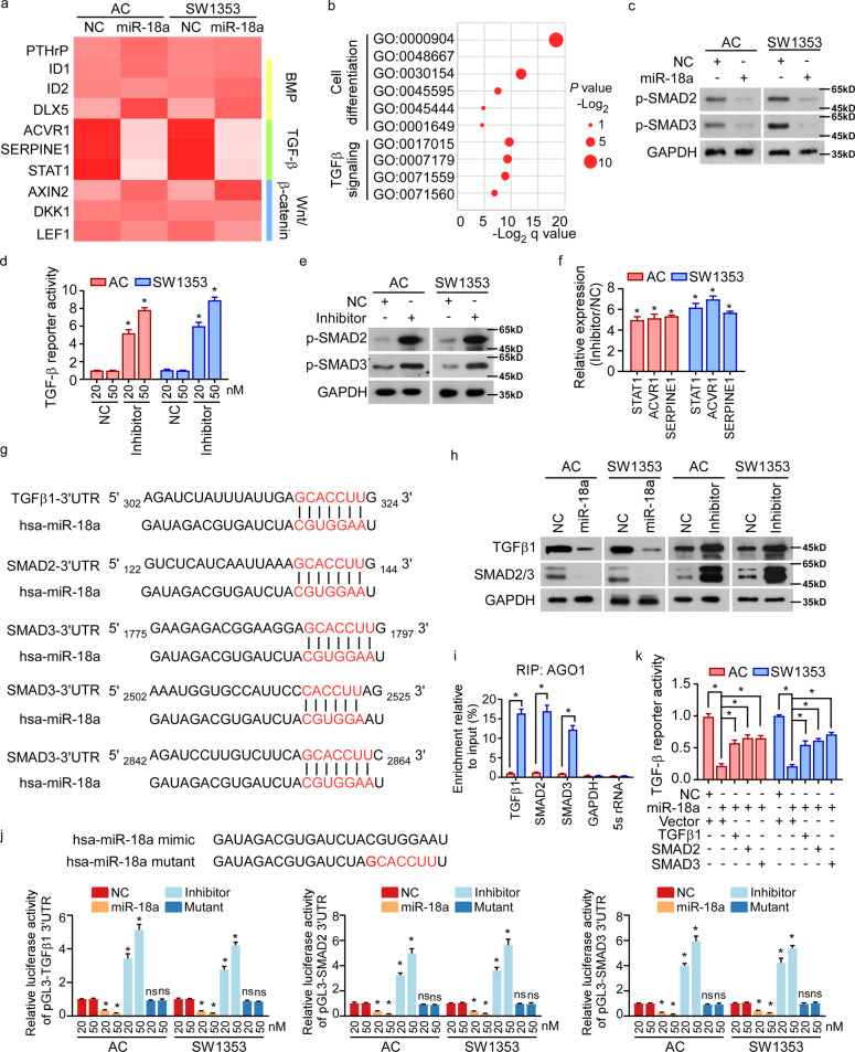
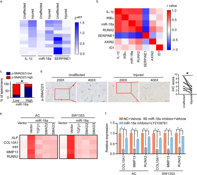
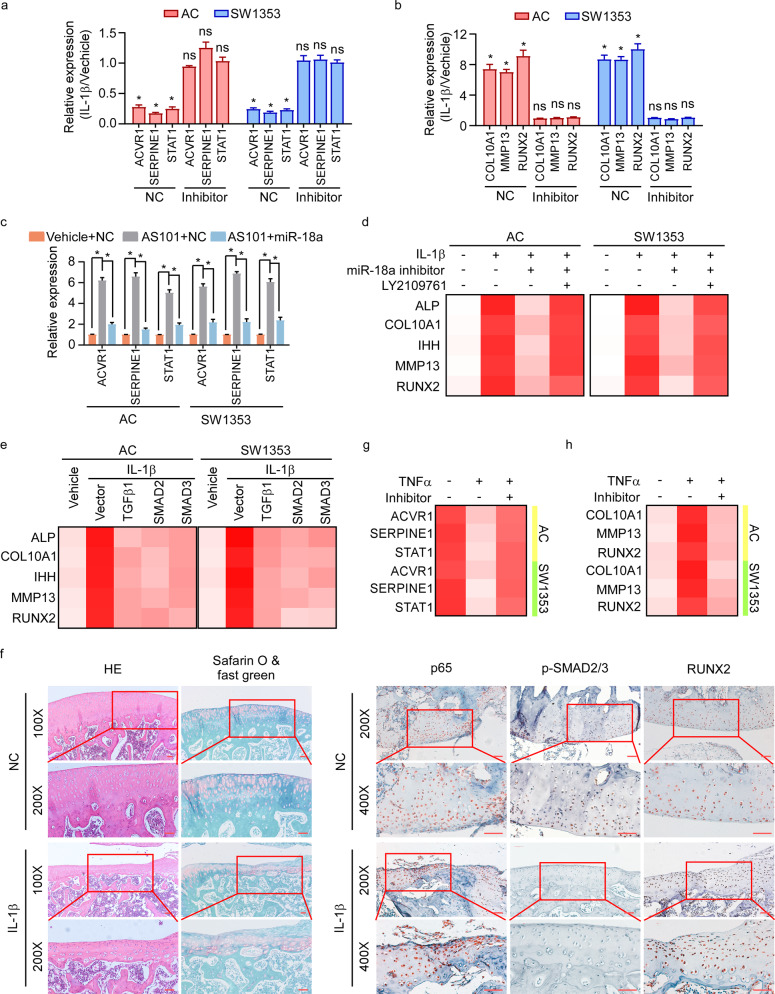
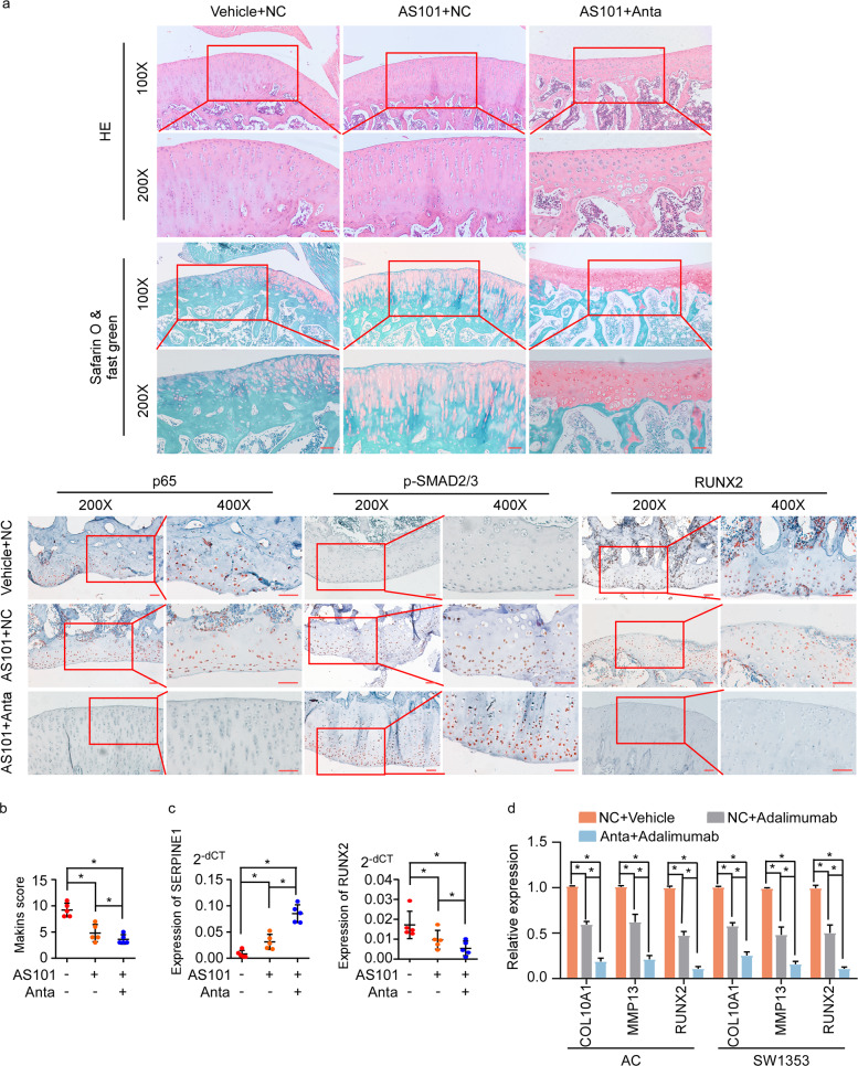
Inflammation participates in the development of OA and targeting inflammatory signaling pathways is a potential strategy for OA treatment. IL-1β is one of the most important inflammatory factors to trigger the activation of NF-κB signaling and accelerate OA progression, whereas OA patients could hardly benefit from inhibiting IL-1β in clinic, suggesting the importance to further explore the details of OA inflammation. We here showed that expression of miR-18a in chondrocytes was specifically induced in response to IL-1β in vitro as well as in rat model of OA during which NF-κB signaling was involved, and that nuclear-translocated p65 directly upregulated miR-18a expression at transcriptional level. Further, increased miR-18a mediated hypertrophy of chondrocytes, resulting in OA degeneration, by targeting TGFβ1, SMAD2, and SMAD3 and subsequently leading to repression of TGF-β signaling. And the level of serum miR-18a was positively correlated to severity of OA. Interestingly, other than IL-1β, pro-inflammation cytokines involving TNFα could also remarkably upregulate miR-18a via activating NF-κB signaling and subsequently induce chondrocytes hypertrophy, suggesting a pivotal central role of miR-18a in inflammatory OA progression. Thus, our study revealed a novel convergence of NF-κB and TGF-β signaling mediated by miR-18a, and a novel mechanism underlying inflammation-regulated OA dependent of NF-κB/miR-18a/TGF-β axis. Notably, in vivo assay showed that targeting miR-18a sensitized OA chondrocytes to IL-1β inhibitor as targeting IL-1β and miR-18a simultaneously had much stronger inhibitory effects on OA progression than suppressing IL-1β alone. Therefore, the diagnostic and therapeutic potentials of miR-18a for OA were also revealed.


Conflict of interest statement

The authors declare no competing interests.

Figures

Fig. 1
Fig. 1. miR-18a was up-regulated in response to IL-1β in OA.
a Schematic representation for identification of significantly altered miRNAs in stimulation of IL-1β in chondrocytes in vitro and in vivo simultaneously. In vitro, RNA was extracted from human articular chondrocytes and SW1353 cells cultured with IL-1β (10 ng/mL) for 2 days. ACLT operation was used to induce knee OA in rats, and the day after surgery vehicle or IL-1β (20 ng/joint) were injected into the rats’ knee joints every 3 days, followed by dissociation of articular cartilage after 4 weeks and extraction of its RNA. qRT-PCR shows the relative expression of miRNAs those had been found to be regulated by IL-1β in rat chondrocytes. b The effect of suppressing NF-κB signaling on the ability of IL-1β to induce expression of miR-18a. c Schematic diagram of potential binding site for p65 in the promoter region of miR-18a (left panel). ChIP enrichment assay shows binding of p65 to the predicted binding site in the promoter region of miR-18a in stimulation of IL-1β, which could be remarkably repressed by JSH23 (right panel). IgG immunoprecipitation was used as a negative control. d ChIP analysis following H3K27ac immunoprecipitation shows the interaction between H3K27ac and the promoter region of miR-18a in response to IL-1β with or without JSH23. IgG immunoprecipitation was used as a negative control. e qRT-PCR to assess the expression of miR-18a in injured articular cartilage of OA patients and the corresponding unaffected cartilage tissues. f Correlation between expression of miR-18a and IL-1β in OA lesions was determined by qRT-PCR and pearson correlation analysis. g Serum miR-18a was assessed in OA patients and healthy volunteer donors. Data in be and g are presented as mean ± SD. *P < 0.05.
Fig. 2
Fig. 2. IL-1β induced-miR-18a accelerates OA progression by promoting chondrocyte hypertrophy.
a Pellet cultures of MSCs expressing vector or miR-18a were induced to undergo chondrogenesis for 14 days and then induced for hypertrophic differentiation for another 14 days. Gross appearance, HE staining, and Alcian blue staining were evaluated. b, c WB b and qRT-PCR c to assess the expression of chondrocyte hypertrophy-related genes when miR-18a was ectopic expressed. d The effect of antagomir of miR-18a (Anta) on hypertrophy of chondrifying MSCs was assessed. e, f Expression of chondrocyte hypertrophy-related genes were determined by WB e and qRT-PCR f in AC and SW1353 cells transfected with inhibitor of miR-18a. g qRT-PCR shows miR-18a level in both injured cartilage and the corresponding paired unaffected cartilage. h The effect of miR-18a antagomir on IL-1β-enhanced hypertrophy of chondrifying MSCs was assessed. i, j Protein i and mRNA j levels of hypertrophy-related genes were analyzed in chondrocytes in response to IL-1β with or without inhibition of miR-18a. k qRT-PCR shows the effect of miR-18a ectopic expression on chondrocyte hypertrophy in cells treated with either AS101 or JSH23. l Expression of IL-1β, miR-18a, and RUNX2 were analyzed in injured cartilage and corresponding unaffected cartilage from OA patients. Scale bar in images of gross appearance in a, d, and h: 1 mm; other scale bars: 200 μm. Data in c, f, and j are presented as mean ± SD. *P < 0.05.
Fig. 3
Fig. 3. miR-18a suppresses TGF-β signaling by targeting TGFβ1, SMAD2, and SMAD3.
a Expression of PTHrP and downstream genes of BMP, TGF-β, and Wnt/β-catenin signaling pathways was analyzed in miR-18a-overexpressed cells. b mRNA array was conducted in human ACs expressing control or miR-18a. And GO-enrichment analysis shows the correlation between expression of miR-18a and cell differentiation-associated signature or TGF-β signaling. c WB shows phosphorylation of SMAD2 and SMAD3 when miR-18a was overexpressed. d Dual-luciferase assays reveal TGF-β signaling activities. e, f Effect of inhibiting miR-18a on activity of TGF-β signaling was analyzed by WB e and qRT-PCR f. g Targetscan tool showing schematic representation of putative binding sites for miR-18a in 3′-UTRs of TGFβ1, SMAD2, and SMAD3. h WB analysis of the protein levels of TGFβ1 and SMAD2/3 in the indicated cells. i By immunoprecipitation against Ago1, RIP analysis reveals the interaction of miR-18a with the 3′-UTRs of TGFβ1, SMAD2, or SMAD3 mRNA to form miRNP complexes. IgG immunoprecipitation, as well as the interaction of miR-18a with GAPDH and 5s rRNA, were used as negative controls. j Luciferase assay of pGL3-TGFβ1-3′-UTR, pGL3-SMAD2-3′-UTR, and pGL3-SMAD3-3′-UTR reporters in the indicated cells, co-transfected with increasing amounts (20 and 50 nM) of the indicated oligonucleotides. The sequence of the miR-18a mutant is shown. k Effects of restored expression of TGFβ1, SMAD2, or SMAD3 in miR-18a-overexpressing cells on luciferase activities of the TGF-β reporter. Data in d, f, i, j, and k are presented as mean ± SD. *P < 0.05.
Fig. 4
Fig. 4. miR-18a mediates chondrocyte hypertrophy via inhibiting TGF-β signaling.
a qRT-PCR to determine the expression of IL-1β, miR-18a, and SERPINE1 in injured cartilage and corresponding paired unaffected cartilage of OA patients. b Pearson’s correlation coefficient between IL-1β, IKBα (NF-κB signaling downstream gene), miR-18a, RUNX2, SERPINE1, AXIN2, and ID1 in injured lesions of OA patients was performed, and the heat map shows r values. c Correlation between p-SMAD2/3 and expression of miR-18a in OA lesions was assessed by IHC, qRT-PCR, and chi-square test. d IHC to determine the level of phosphorylation of SMAD2/3 in unaffected and injured articular cartilage. Scale bar: 50 μm. e Effects of restored expression of TGFβ1, SMAD2, or SMAD3 in miR-18a-overexpressing cells on expression of hypertrophy-related genes. f qRT-PCR exhibits the expression of hypertrophy-related genes in AC and SW1353 cells expressing inhibitor of miR-18a with or without LY2109761. Data in f are presented as mean ± SD. *P < 0.05.
Fig. 5
Fig. 5. IL-1β-mediated hypertrophy depends on miR-18a-induced suppression of TGF-β signaling.
a Effects of IL-1β on activation of TGF-β signaling in AC and SW1353 cells expressing NC or miR-18a inhibitor were assessed. b Expression of hypertrophy-related genes was analyzed in NC- or miR-18a inhibitor-expressed cells in response to IL-1β. c TGF-β signaling downstream genes expression was analyzed in IL-1β-suppressed cells with or without miR-18a re-expression. d Effects of LY2109761 on chondrocyte hypertrophy in cells with IL-1β treatment and miR-18a inhibitor expression were determined. e qRT-PCR shows the expression of hypertrophy-related genes when TGFβ1, SMAD2, or SMAD3 was restored expressed in cells with IL-1β stimulation. f Activity of NF-κB and TGF-β signaling, and expression of RUNX2 were determined in rat model of OA in Fig. 1a. Scale bar: 50 μm. g, h Activity of TGF-β signaling and the expression of hypertrophic genes in chondrocytes were assessed in cells in response to TNFα with or without inhibition of miR-18a. Data in ac are presented as mean ± SD. *P < 0.05.
Fig. 6
Fig. 6. Targeting miR-18a sensitized chondrocytes in OA lesions to anticytokine drugs.
a AS101 (1 mg/kg) was intraperitoneal injected, and 10 μL antagomir of miR-18a (250 nM) were intra-articular injected in rat model of knee OA every 3 days, followed by dissociation of knee joint after 4 weeks. HE staining, Safarin O, and fast green staining, and IHC of p65, p-SMAD2/3, and RUNX2 were conducted. Scale bar: 50 μm. b Mankin’s score was assessed. c Expression of SERPINE1 and RUNX2 in articular cartilage from OA rats with administration of AS101 or combination of AS101 and miR-18a antagomir. d qRT-PCR to determine the expression of hypertrophy-related genes in human ACs and SW1353 cells with treatment of adalimumab or combination of adalimumab and miR-18a antagomir. Data in bd are presented as mean ± SD. *P < 0.05.

References

    1. Robinson WH, et al. Low-grade inflammation as a key mediator of the pathogenesis of osteoarthritis. Nat. Rev. Rheumatol. 2016;12:580–592. doi: 10.1038/nrrheum.2016.136. - DOI - PMC - PubMed
    1. Scanzello CR, Goldring SR. The role of synovitis in osteoarthritis pathogenesis. Bone. 2012;51:249–257. doi: 10.1016/j.bone.2012.02.012. - DOI - PMC - PubMed
    1. Page Thomas DP, King B, Stephens T, Dingle JT. In vivo studies of cartilage regeneration after damage induced by catabolin/interleukin-1. Ann. Rheum. Dis. 1991;50:75–80. doi: 10.1136/ard.50.2.75. - DOI - PMC - PubMed
    1. Henderson B, Pettipher ER. Arthritogenic actions of recombinant IL-1 and tumour necrosis factor alpha in the rabbit: evidence for synergistic interactions between cytokines in vivo. Clin. Exp. Immunol. 1989;75:306–310. - PMC - PubMed
    1. Merz D, Liu R, Johnson K, Terkeltaub R. IL-8/CXCL8 and growth-related oncogene alpha/CXCL1 induce chondrocyte hypertrophic differentiation. J. Immunol. 2003;171:4406–4415. doi: 10.4049/jimmunol.171.8.4406. - DOI - PubMed

Publication types

MeSH terms