. 2020 Nov 3;10(1):18946.
doi: 10.1038/s41598-020-75873-1.

Apoptosis, the only cell death pathway that can be measured in human diploid dermal fibroblasts following lethal UVB irradiation

Affiliations

Apoptosis, the only cell death pathway that can be measured in human diploid dermal fibroblasts following lethal UVB irradiation

Anne-Sophie Gary et al. Sci Rep. .

Abstract

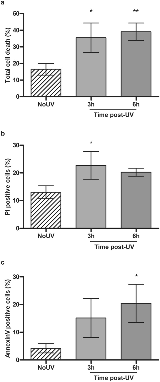
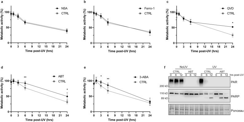
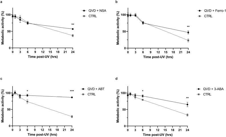
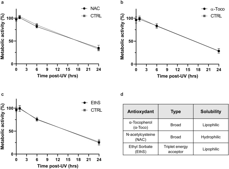
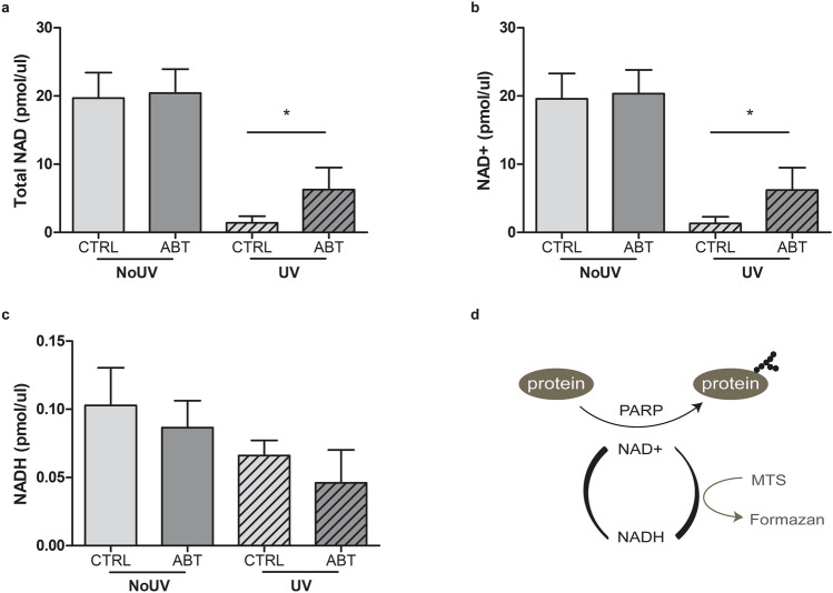
Ultraviolet radiation (UVR) is a major environmental genotoxic agent. In skin, it can lead to the formation of mutagenic DNA damage. Several mechanisms are in place to prevent the conversion of these DNA damage into skin cancer-driver mutations. An important mutation prevention mechanism is the programmed cell death, which can safely dispose of the damaged cells. Apoptosis is the most studied and best characterised programmed cell death, but an increasing amount of new cell death pathways are emerging. Using different pharmacological cell death inhibitors and antioxidants, we have evaluated the implication of apoptosis, necroptosis, ferroptosis and parthanatos in UVB-induced cell death in human diploid dermal fibroblasts. Our results show that apoptosis is the only known cell death mechanism induced by UVB irradiation in fibroblasts. We also showed that lethal UVB irradiation induces a PARP-dependent drastic loss of cellular metabolic activity caused by an overused of NAD+.


Conflict of interest statement

The authors declare no competing interests.

Figures

Figure 1
Figure 1
UVB-induced early cell death in primary human fibroblasts. Using Annexin V-FITC and PI labelling, we assessed cell death in fibroblasts at 3 h and 6 h following an irradiation with 20 kJ/m2 UVB. The fluorescence signal in each cell was assessed by FACS. (a) Total cell death is the addition of Annexin V+/PI− cells, Annexin V+/PI+ cells and Annexin V−/PI+ cells. (b) PI positive cells is composed of PI+/Annexin V+ or − cells. (c) Annexin V positive cells are Annexin V+ and PI−. NoUV are unirradiated control cells. UVB irradiation of fibroblasts leads to cell death as early as 3 h. Percentage is representative of a population of at least 8000 cells. N = 3, *p-value < 0.05.
Figure 2
Figure 2
Cellular metabolic activity of UVB-irradiated primary human fibroblasts treated with different cell death inhibitors. Prior to UVB irradiation, fibroblasts were incubated 30 min with different cell death inhibitors. Cells were then irradiated in PBS using a lethal UVB dose (30 kJ/m2). The different cell death inhibitors used were: (a) The necroptosis inhibitor Necrosulfonamide (NSA, 2 μM), (b) the ferroptosis inhibitor Ferrostatin-1 (Ferro-1, 5 μM), (c) the broad caspase inhibitor Q-VD-OPh (QVD, 20 μM), (d) the PARP inhibitor ABT888 (ABT, 20 μM) or (e) the PARP inhibitor 3-Aminobenzamide (3-ABA, 0.5 mM). Cellular metabolic activity was assessed at different time points post-UVB irradiation (0, 1, 3, 6, 24 h) using MTS assay. Irradiated cells were normalised on unirradiated cells of the same condition. NSA and Ferro-1 had virtually no effect on cellular metabolism. However, QVD significantly prevent UVB-induced cellular metabolic activity loss at 24 h, ABT at 6 and 24 h and 3-ABA at 3 and 6 h. N = 4. *p-value < 0.05, **p-value < 0.01, ***p-value < 0.001. (f) Efficiency of ABT888 was confirmed by Western Blot, showing an inhibition of PAR formation without abolition of apoptotic PARP cleavage.
Figure 3
Figure 3
UVB induces two independent reduction of cellular metabolic activity in primary human fibroblasts. Prior to UVB irradiation, fibroblasts were incubated 30 min with different combinations of cell death inhibitors. Cells were then irradiated in PBS using a lethal UVB dose (30 kJ/m2). The combinations used were: (a) Necroptosis inhibitor Necrosulfonamide (NSA, 2 μM) and caspase inhibitor Q-VD-OPh (QVD, 20 μM), (b) ferroptosis inhibitor Ferrostatin-1 (Ferro-1, 5 μM) and caspase inhibitor Q-VD-OPh (QVD, 20 μM), (c) PARP inhibitor ABT888 (ABT, 20 μM) and caspase inhibitor Q-VD-OPh (QVD, 20 μM), (d) PARP inhibitor 3-Aminobenzamide (3-ABA, 0.5 mM) and caspase inhibitor Q-VD-OPh (QVD, 20 μM). Cellular metabolic activity was assessed at different time points post UVB irradiation (0, 1, 3, 6, 24 h) using MTS assay. Irradiated cells were normalised on unirradiated cells of the same condition. NSA and Ferro-1 had no effect on cellular metabolic activity loss prevention caused by QVD. However, an additive effect of QVD and PARP inhibitors (ABT and 3-ABA) on prevention of cellular metabolic activity loss caused by UVB could be described. N = 4. *p-value < 0.05, **p-value < 0.01, ***p-value < 0.001.
Figure 4
Figure 4
UVB-induced cell death is independent of oxidation in primary human fibroblasts. Prior to UVB irradiation, fibroblasts were incubated 30 min with different antioxidants. Cells were then irradiated in PBS using a lethal UVB dose (30 kJ/m2). The different antioxidant used were: (a) Broad-spectrum oxidation inhibitor N-acetylcysteine (NAC, 2.5 mM), (b) broad-spectrum oxidation inhibitor α-Tocopherol (α-Toco, 10 μM), (c) triplet energy acceptor Ethyl Sorbate (EthS, 5 μg/μl). Cellular metabolic activity was assessed at different time points post UVB exposition (0, 1, 6, 24 h) using a MTS assay. Irradiated cells were normalised on unirradiated cells of the same condition. None of the antioxidant tested had an effect on UVB-induced cellular metabolic activity loss. N = 4. (d) Table of antioxidants used, their type and solubility.
Figure 5
Figure 5
The UVB-induced total NAD and NAD+ pool decrease can partially be restored using PARP inhibitor. Prior to UVB irradiation, fibroblasts were incubated 30 min with or without ABT888 (ABT, 20 μM). Cells were then UVB irradiated with 20 kJ/m2 (UV) or not (NoUV) in PBS. 6 h post exposition, cells were harvested to quantify NAD/NADH. Same amount of proteins was taken to compare the different conditions, i.e. NoUV CTRL, NoUV ABT, UV CTRL and UV ABT. (a) Concentration of Total NAD and (b) concentration of NADH were measured. Concentration of NAD+ was deduced by subtracting NADH to Total NAD in (c). We found that UVB irradiation significantly reduces total NAD and NAD+, but the use of PARP inhibitor ABT significantly prevent this loss. N = 4, *p-value < 0.05. (d) Schematic representation of the link between NAD+ consumption, PARP1 and the MTS assay.
Figure 6
Figure 6
Parthanatos is not induced by UVB in primary human fibroblasts. Fibroblasts were UVB irradiated with 20 kJ/m2 (UV) or not (NoUV). At different time points after exposition, cells were harvested (0, 1, 3, 6, 9, 12 h) and proteins from mitochondria, nucleus and cytoplasm were separated. Western Blot against AIF using protein fractions of (a) mitochondria, (b) nucleus and (c) cytoplasm showed no translocation of the protein from mitochondria to nucleus. Controls of fractionation were measured by simultaneously scanning the membranes from the 3 fractions. Nuclear, cytosolic and mitochondrial controls are Lamin A/C, Tubulin and AIF, respectively. Ponceau staining has been used as loading control. (d) Prior to UVB irradiation at 30 kJ/m2, cells were incubated 30 min with ABT888 (ABT, 20 μM), Q-VD-OPh (QVD, 20 μM) or control media (CTRL). Immediately after UVB exposure cells were incubated with corresponding inhibitor and CellTOX dye. CellTOX fluorescence, proportional to cell death, was measure at different time points (0, 6, 12 and 24 h). Fluorescence from unirradiated cells (background) was subtracted to irradiated cells for the same condition. The apoptosis inhibitor QVD significantly prevents UVB-induced cell death at 12 and 24 h. However, the PARP inhibitor ABT had no effect on UVB-induced cell death. SEM, N = 4, *p-value < 0.05.

Similar articles

Cited by

References

    1. Rastogi RP, Richa KA, Tyagi MB, Sinha RP. Molecular mechanisms of ultraviolet radiation-induced DNA damage and repair. J. Nucleic Acids. 2010;2010:592980. doi: 10.4061/2010/592980. - DOI - PMC - PubMed
    1. Kerr JB, Fioletov VE. Surface ultraviolet radiation. Atmos. Ocean. 2008;46:159–184. doi: 10.3137/ao.460108. - DOI
    1. Iwasaki K, Izawa M, Mihara M. UV-induced apoptosis in rat skin. J. Dermatol. Sci. 1996;12:31–35. doi: 10.1016/0923-1811(95)00461-0. - DOI - PubMed
    1. Courdavault S, et al. Repair of the three main types of bipyrimidine DNA photoproducts in human keratinocytes exposed to UVB and UVA radiations. DNA Repair (Amst). 2005;4:836–844. doi: 10.1016/j.dnarep.2005.05.001. - DOI - PubMed
    1. Wickert H, et al. Differential induction of proto-oncogene expression and cell death in ocular tissues following ultraviolet irradiation of the rat eye. Br. J. Ophthalmol. 1999;83:225–230. doi: 10.1136/bjo.83.2.225. - DOI - PMC - PubMed

Publication types

MeSH terms

Substances