. 2020 Nov 3;10(1):18897.
doi: 10.1038/s41598-020-75731-0.

A comprehensive diagnostic approach combining phylogenetic disease bracketing and CT imaging reveals osteomyelitis in a Tyrannosaurus rex

Affiliations

A comprehensive diagnostic approach combining phylogenetic disease bracketing and CT imaging reveals osteomyelitis in a Tyrannosaurus rex

C A Hamm et al. Sci Rep. .

Abstract

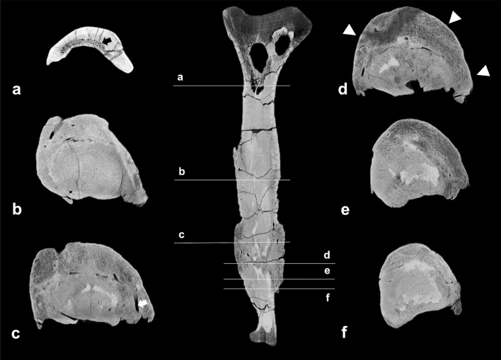
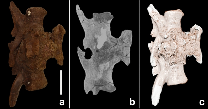
Traditional palaeontological techniques of disease characterisation are limited to the analysis of osseous fossils, requiring several lines of evidence to support diagnoses. This study presents a novel stepwise concept for comprehensive diagnosis of pathologies in fossils by computed tomography imaging for morphological assessment combined with likelihood estimation based on systematic phylogenetic disease bracketing. This approach was applied to characterise pathologies of the left fibula and fused caudal vertebrae of the non-avian dinosaur Tyrannosaurus rex. Initial morphological assessment narrowed the differential diagnosis to neoplasia or infection. Subsequent data review from phylogenetically closely related species at the clade level revealed neoplasia rates as low as 3.1% and 1.8%, while infectious-disease rates were 32.0% and 53.9% in extant dinosaurs (birds) and non-avian reptiles, respectively. Furthermore, the survey of literature revealed that within the phylogenetic disease bracket the oldest case of bone infection (osteomyelitis) was identified in the mandible of a 275-million-year-old captorhinid eureptile Labidosaurus. These findings demonstrate low probability of a neoplastic aetiology of the examined pathologies in the Tyrannosaurus rex and in turn, suggest that they correspond to multiple foci of osteomyelitis.


Conflict of interest statement

The authors declare no competing interests.

Figures

Figure 1
Figure 1
Tyrannosaurus rex specimen FMNH PR2081 and gross appearance of the fused caudals and left fibula. (a,b) Illustration of the articulated skeleton of the Tyrannosaurus rex ‘Sue’. The bones under investigation in this study, the left fibula and fused caudal vertebrae c26 and c27, are highlighted in red. (cf) The readily apparent circumferential rugose bone formation of the fused caudals is indicated by arrowheads. (g,h) Fibula with magnification of pathological changes; arrowheads indicate the rugose surface. (Copyright Scott Hartman, 2019. Modified and used with permission).
Figure 2
Figure 2
Fibula. (a) Gross appearance; (b,c) sagittal cross-sectional CT images; (d) 3D reconstruction based on CT images. The sagittal cross-sectional CT images demonstrate (i) increased bone thickness, limited to the two horizontal white bars; (ii) decreased bone density in the area of the active focus of the infection (arrowheads); (iii) tubular canals in the periosteal bone formation (arrows). The scale bar represents 10 cm.
Figure 3
Figure 3
Cross-sectional CT images of the fibula at different levels of the bone, demonstrating the changes of bone architecture throughout the bone. Arrowheads indicate the pronounced periosteal bone formation. The white arrow indicates one of the tubular canals of the periosteal bone formation. The black arrow indicates the trabecular bone in the proximal part of the fibula.
Figure 4
Figure 4
Fused caudals. (a) Gross appearance; (b) sagittal cross-sectional CT image; (c) 3D reconstruction based on CT images. The scale bar represents 10 cm.
Figure 5
Figure 5
Cross-sectional images of fused caudals. The pathological changes are recognisable on the coronal (a), sagittal (b) and axial (c) plane. The axial plane demonstrates the cross-section of the bone on the level of the blue line, indicated in the sagittal plane. The black asterisks (*) indicates areas with increased density within the lesion, presumably due to sediment enclosures; the white arrow indicates decreased bone density in the area between the haemal arch and intervertebral articulation, suggesting an active focus of infection.
Figure 6
Figure 6
Neoplastic and infectious diseases in the dinosaur cladogram. Simplified non-avian dinosaur family tree showing the distribution of infectious (orange) and neoplastic diseases (blue) as derived from the literature analysis (numbers indicate the respective references). For Aves and non-avian reptiles, the prevalences of the respective diseases are given as percentages and derived from the pooled literature analysis given in Tables 1 and 2. The specific neoplastic disease rate for the non-avian reptile taxons testudines, lepidosaurs and crocodylians are derived from the pooled literature analysis given in the Supplementary Table 2,,,,–,,,–,,,,,,,,,,,,–.

Similar articles

Cited by

References

    1. Hanna RR. Multiple injury and infection in a sub-adult theropod dinosaur Allosaurus fragilis with comparisons to allosaur pathology in the Cleveland-Lloyd Dinosaur Quarry collection. J. Vertebr. Paleontol. 2002;22:76–90. doi: 10.1671/0272-4634(2002)022[0076:miaiia]2.0.co;2. - DOI
    1. Taylor LH, Latham SM, Mark E. Risk factors for human disease emergence. Philos. Trans. R. Soc. B Biol. Sci. 2001;356:983–989. doi: 10.1098/rstb.2001.0888. - DOI - PMC - PubMed
    1. Tanke, D. H. & Rothschild, B. M. Dinosores: An annotated bibliography of dinosaur paleopathology and related topics—1838–2001: Bulletin 20. Vol. 20 1–97 (New Mexico Museum of Natural History and Science, 2002).
    1. Rothschild B. Scientifically rigorous reptile and amphibian osseous pathology: Lessons for forensic herpetology from comparative and paleo-pathology. Appl. Herpetol. 2009;6:47–79. doi: 10.1163/157075409x413842. - DOI
    1. Rothschild BM, Tanke D. Paleopathology of vertebrates: Insights to lifestyle and health in the geological record. Geosci. Can. 1992;19:73–82.

Publication types