

Transcriptomic profiling of feline teeth highlights the role of matrix metalloproteinase 9 (MMP9) in tooth resorption
- PMID: 33144645
- PMCID: PMC7641192
- DOI: 10.1038/s41598-020-75998-3
Transcriptomic profiling of feline teeth highlights the role of matrix metalloproteinase 9 (MMP9) in tooth resorption
Abstract
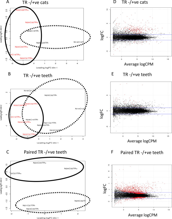
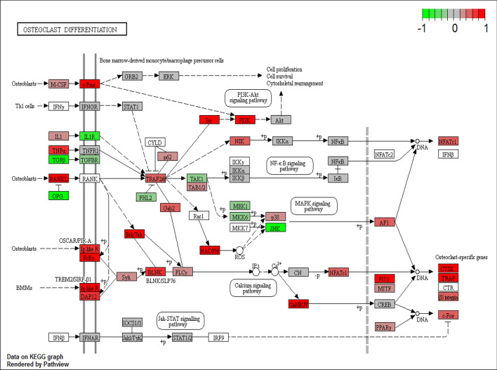
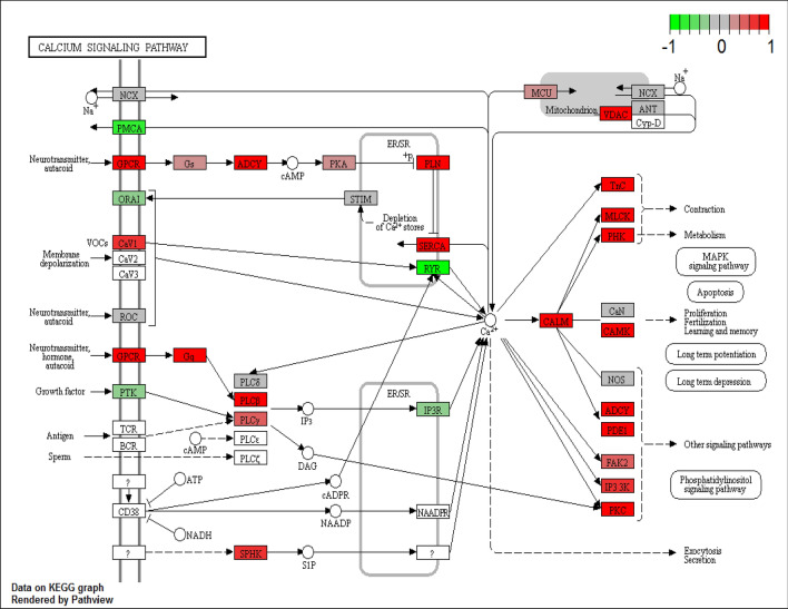
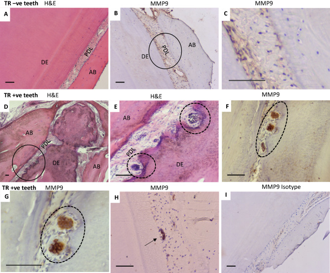
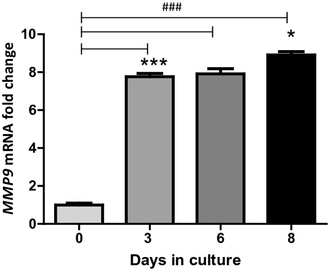
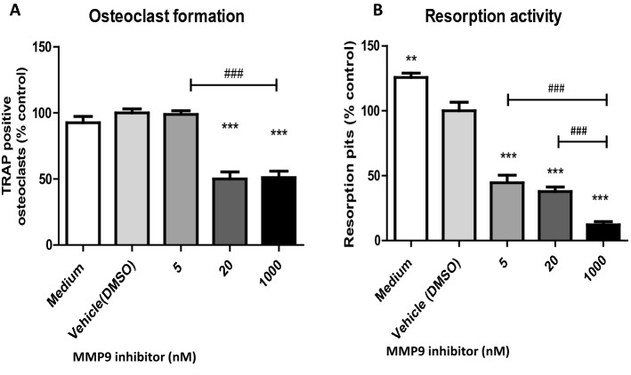
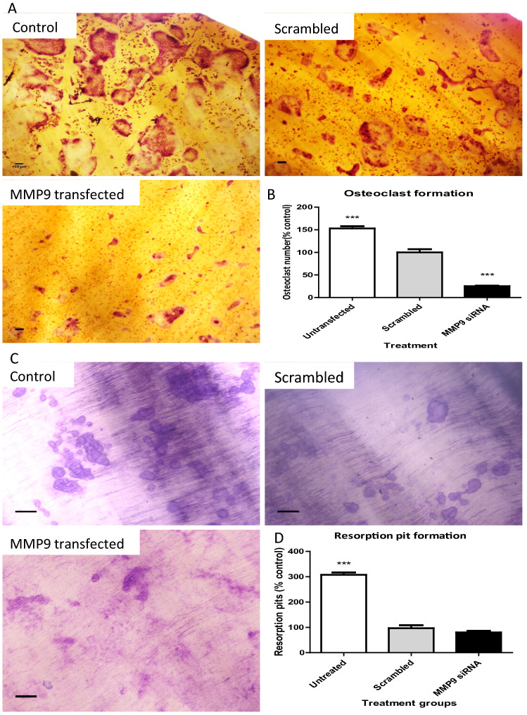
Tooth resorption (TR) in domestic cats is a common and painful disease characterised by the loss of mineralised tissues from the tooth. Due to its progressive nature and unclear aetiology the only treatment currently available is to extract affected teeth. To gain insight into TR pathogenesis, we characterised the transcriptomic changes involved in feline TR by sequencing RNA extracted from 14 teeth (7 with and 7 without signs of resorption) collected from 11 cats. A paired comparison of teeth from the same cat with and without signs of resorption identified 1,732 differentially expressed genes, many of which were characteristic of osteoclast activity and differentiation, in particular matrix metalloproteinase 9 (MMP9). MMP9 expression was confirmed by qPCR and immunocytochemistry of odontoclasts located in TR lesions. A hydroxamate-based MMP9 inhibitor reduced both osteoclast formation and resorption activity while siRNA targeting MMP9 also inhibited osteoclast differentiation although had little effect on resorption activity. Overall, these results suggest that increased MMP9 expression is involved in the progress of TR pathogenesis and that MMP9 may be a potential therapeutic target in feline TR.
Conflict of interest statement
The authors declare no competing interests.
Figures
Similar articles
-
Prevalence of Tooth Resorptive Lesions in 120 Feline Dental Patients in Israel.J Vet Dent. 2025 Mar;42(2):114-117. doi: 10.1177/08987564231226082. Epub 2024 Jan 31. J Vet Dent. 2025. PMID: 38295354
-
A large case-control study indicates a breed-specific predisposition to feline tooth resorption.Vet J. 2024 Jun;305:106133. doi: 10.1016/j.tvjl.2024.106133. Epub 2024 May 11. Vet J. 2024. PMID: 38740176
-
Expression of matrix metalloproteinase-9 mRNA and protein during deciduous tooth resorption in bovine odontoclasts.Bone. 2002 Oct;31(4):472-8. doi: 10.1016/s8756-3282(02)00856-6. Bone. 2002. PMID: 12398942
-
Tooth resorption in cats: pathophysiology and treatment options.J Feline Med Surg. 2015 Jan;17(1):37-43. doi: 10.1177/1098612X14560098. J Feline Med Surg. 2015. PMID: 25527492 Free PMC article. Review.
-
Update on the etiology of tooth resorption in domestic cats.Vet Clin North Am Small Anim Pract. 2005 Jul;35(4):913-42, vii. doi: 10.1016/j.cvsm.2005.03.006. Vet Clin North Am Small Anim Pract. 2005. PMID: 15979519 Review.
Cited by
-
Deciphering 3D periodontal fibroblast-macrophage crosstalk in bioactive nanoparticle-guided immunomodulation for treating traumatic dental avulsion.Bioact Mater. 2024 Jul 31;41:400-412. doi: 10.1016/j.bioactmat.2024.07.017. eCollection 2024 Nov. Bioact Mater. 2024. PMID: 39184829 Free PMC article.
-
CX3CL1 promotes M1 macrophage polarization and osteoclast differentiation through NF-κB signaling pathway in ankylosing spondylitis in vitro.J Transl Med. 2023 Aug 25;21(1):573. doi: 10.1186/s12967-023-04449-0. J Transl Med. 2023. PMID: 37626378 Free PMC article.
-
Resilience of the replacing dentition in adult reptiles.Dev Biol. 2024 Dec;516:71-81. doi: 10.1016/j.ydbio.2024.07.013. Epub 2024 Jul 24. Dev Biol. 2024. PMID: 39059678 Free PMC article. Review.
-
Evaluation of Feline Permanent Canine Tooth Mineral Density Using Micro-Computed Tomography.Vet Sci. 2023 Mar 12;10(3):217. doi: 10.3390/vetsci10030217. Vet Sci. 2023. PMID: 36977256 Free PMC article.
-
Exosome: Function and Application in Inflammatory Bone Diseases.Oxid Med Cell Longev. 2021 Aug 31;2021:6324912. doi: 10.1155/2021/6324912. eCollection 2021. Oxid Med Cell Longev. 2021. Retraction in: Oxid Med Cell Longev. 2024 Jan 9;2024:9806854. doi: 10.1155/2024/9806854. PMID: 34504641 Free PMC article. Retracted. Review.
References
Publication types
MeSH terms
Substances
Grants and funding
LinkOut - more resources
Full Text Sources
Miscellaneous
