Review
. 2020 Sep;12(9):5128-5139.
doi: 10.21037/jtd.2019.08.37.

Thyroid cancer diagnosis in the era of precision imaging

Affiliations
Review

Thyroid cancer diagnosis in the era of precision imaging

Kimberley-Jane Bonjoc et al. J Thorac Dis. 2020 Sep.

Abstract

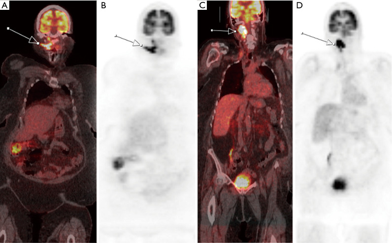
Thyroid cancer affects 1.3% of the population with increasing rates of incidence over the last decade (approximately 2% per year). Although the overall prognosis is good in the differentiated subtypes, there has been a slow but steady increase in rate of deaths associated with thyroid cancer (approximately 0.7% per year over the last decade). Thyroid cancer is usually detected when: (I) patients feel a lump in the neck; (II) a routine clinical exam is performed; (III) an incidental thyroid nodule is identified on diagnostic imaging (e.g., CT neck or chest, carotid ultrasound, PET scan acquired for non-thyroid pathology). Identification of suspicious thyroid nodules results in further diagnostic work-up including laboratory assessment, further imaging, and biopsy. Accurate diagnosis is required for clinical staging and optimal patient treatment design. In this review, we aim to discuss utility of various imaging modalities and their role in thyroid cancer diagnosis and management. Additionally, we aim to highlight emerging diagnostic techniques that aim to improve diagnostic specificity and accuracy in thyroid cancer, thus paving way for precision medicine.

Keywords: Positron emission tomography/computed tomography (PET/CT); cancer; magnetic resonance imaging (MRI); thyroid; ultrasound.


Conflict of interest statement

Conflicts of Interest: All authors have completed the ICMJE uniform disclosure form (available at http://dx.doi.org/10.21037/jtd.2019.08.37). The series “Role of Precision Imaging in Thoracic Disease” was commissioned by the editorial office without any funding or sponsorship. AC served as the unpaid Guest Editor for the series. The other authors have no other conflicts of interest to declare.

Figures

Figure 1
Figure 1
A 60-year-old female’s transverse and longitudinal ultrasound presents multiple micropapillary carcinomas with microcalcification and increased vascularity.
Figure 2
Figure 2
A 82-year-old female diagnosed with metastatic medullary and classical papillary thyroid carcinoma. Transverse (left) and longitudinal (right) ultrasound displays thyroid lesion with microcalcifications indicated by white arrows.
Figure 3
Figure 3
A 39-year-old male patient diagnosed with metastatic papillary thyroid carcinoma. Patient presents 131-Iodine uptake in I-SPECT scan, indicating follicular adenoma.
Figure 4
Figure 4
Clinical work-up for incidental thyroids identified on computed tomography (24).
Figure 5
Figure 5
Coronal (left) and sagittal (right) CT of a 39-year-old male patient diagnosed with metastatic papillary thyroid carcinoma. Patient presents a heterogeneously enhancing thyroid nodule that is eroding through the thyroid capsule (white arrows).
Figure 6
Figure 6
A 66-year-old female diagnosed with poorly differentiated thyroid carcinoma. FDG PET/CT demonstrates increased FDG uptake in the thyroid nodule (white arrow) Additionally, there is FDG avid metastasis to ipsilateral cervical chain lymph node (blue arrow). PET/CT, positron emission tomography/computed tomography; FDG, fluorodeoxyglucose.
Figure 7
Figure 7
A 74-year-old female diagnosed with anaplastic thyroid carcinoma. PET/CT scan shows increased FDG uptake within an enlarged thyroid with irregular margins (white arrow) encasing the trachea. PET/CT, positron emission tomography/computed tomography; FDG, fluorodeoxyglucose.
Figure 8
Figure 8
Sagittal (left) and axial (right) T1-post contrast MRI of a 37-year-old female diagnosed with papillary thyroid carcinoma. T1-post-contrast MRI demonstrates heterogeneously enhancing nodule in the anterior aspect of left thyroid lobe (arrows) with well-circumscribed borders without evidence of ipsilateral adenoapthy or tracheal invasion. MRI, magnetic resonance imaging.
Figure 9
Figure 9
A 79-year-old female diagnosed with metastatic Hurthle cell carcinoma presents radioactive iodine (RAI)-resistance. Initial FDG PET/CT shows increased metabolic activity in the recurrent metastasic right level II/III cervical chain lymph nodes (white arrows) in 2016. Six months after RAI therapy, follow-up FDG PET/CT in 2017 shows increased size and metabolic activity in the right level II/III cervical chain lymph nodes, indicating RAI-resistance. PET/CT, positron emission tomography/computed tomography; FDG, fluorodeoxyglucose; RAI, radioactive iodine.

References

    1. Shore SL. Thyroid Cancer. In: Vinjamuri S, editor. PET/CT in Thyroid Cancer. Cham: Springer International Publishing; 2018. p. 1-7.
    1. Shore SL. Thyroid Cancer Pathology. In: Vinjamuri S, editor. PET/CT in Thyroid Cancer. Cham: Springer International Publishing; 2018. p. 9-13.
    1. Haymart MR, Banerjee M, Reyes-Gastelum D, et al. Thyroid Ultrasound and the Increase in Diagnosis of Low-Risk Thyroid Cancer. J Clin Endocrinol Metab 2019;104:785-92. 10.1210/jc.2018-01933 - DOI - PMC - PubMed
    1. Kitahara CM, Sosa JA. The changing incidence of thyroid cancer. Nat Rev Endocrinol 2016;12:646-53. 10.1038/nrendo.2016.110 - DOI - PMC - PubMed
    1. Lim H, Devesa SS, Sosa JA, et al. Trends in Thyroid Cancer Incidence and Mortality in the United States, 1974-2013. JAMA 2017;317:1338-48. 10.1001/jama.2017.2719 - DOI - PMC - PubMed