. 2020 Oct 22:7:101115.
doi: 10.1016/j.mex.2020.101115. eCollection 2020.

Formulation of PLGA nanoparticles containing short cationic peptide nucleic acids

Affiliations

Formulation of PLGA nanoparticles containing short cationic peptide nucleic acids

Shipra Malik et al. MethodsX. .

Abstract

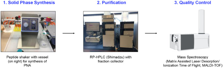
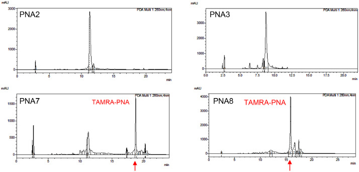
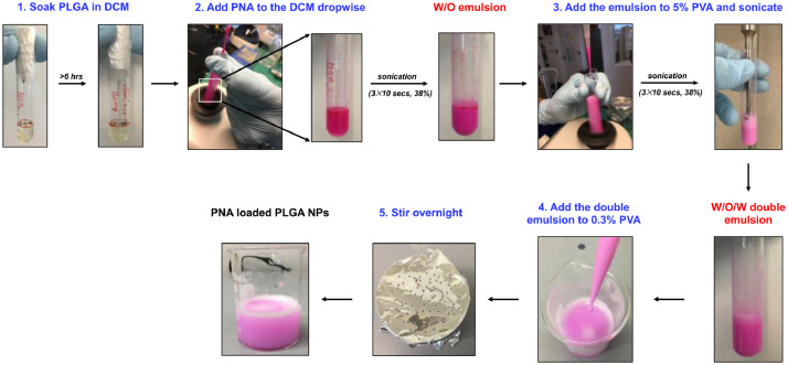
Peptide nucleic acids (PNAs) have emerged as one of the most versatile tools with a wide range of biomedical applications including antisense, antimiR, antigene, as well as site-specific gene editing. The application and potential of PNAs has been limited due to low solubility and poor cellular uptake. Several strategies have been employed to overcome the aforementioned challenges like conjugation to cationic peptides or nanotechnology to achieve superior transfection efficiency ex vivo and in vivo. Here, we report a detailed procedure optimized in our lab for synthesis of short cationic PNA probes, which exhibit high purity and yield in comparison to full-length PNA oligomers. We also provide step-by-step details of encapsulating short cationic PNA probes in poly (lactic-co-glycolic acid) nanoparticles by double emulsion solvent evaporation technique. 1.Detailed procedure for synthesis of short cationic PNAs with or without fluorophore (dye) conjugation while ensuring high yield and purity.2.Step-by-step details for encapsulation of short cationic PNAs in PLGA nanoparticles via double emulsion solvent evaporation technique.

Keywords: AntimiR; Nanoformulations; Nucleic acids.


Conflict of interest statement

The authors declare that they have no known competing financial interests or personal relationships that could have appeared to influence the work reported in this paper.

Figures

Image, graphical abstract
Graphical abstract
Fig 1
Fig. 1
Major steps for synthesizing peptide nucleic acids (PNAs). PNAs are synthesized on a solid support following solid-phase synthesis (Step 1) protocols. After the completion of PNA synthesis, it is cleaved from the solid support and purified via reverse phase high performance liquid chromatography (RP-HPLC) (Step 2) and the mass of the purified PNA (Step 3) is determined via Matrix Assisted Laser Desorption/ Ionization Time of Flight (MALDI-TOF) spectroscopy.
Fig2
Fig. 2
(A) Chemical structure of PNA monomers including Adenine (A), Guanine (G), Cytosine (C) and Thymine (T) used in PNA synthesis. (B) Work flow representing the steps in PNA synthesis. Here, MBHA resin acts as the solid support for PNA synthesis, and first conjugated with an amino acid, Arginine (R) or lysine (K) (Step 1). After conjugation of amino acid to the resin, PNA monomers are conjugated based on the desired sequence (Step 2). This step is divided into three parts including deprotection, coupling, and capping reactions where kaiser test is performed twice for each monomer addition to confirm the completion of reactions. During deprotection, protecting group (Boc) is cleaved resulting in exposure of free amines and is indicated by a blue kaiser test. Next, coupling solution containing the PNA monomer is added and reaction is allowed to continue for 2 h. The completion of coupling reaction is confirmed by a yellow kaiser test, and if the kaiser is blue, coupling reaction is performed again until the kaiser shows yellow. After the completion of coupling, capping is performed for 2 min and synthesis proceeds to deprotection reaction to add the next PNA monomer. These steps of deprotection, coupling and capping are repeated until the entire length of PNA is synthesized. Next, the PNA sequence synthesized is cleaved from the resin (Step 3) and purified by reverse phase HPLC (RP-HPLC) for further use.
Fig 3
Fig. 3
HPLC chromatograms of short cationic crude PNAs. PNA2 is lysine (K) conjugated short PNA and PNA7 is TAMRA conjugated version of PNA2. PNA3 is arginine (R) conjugated short PNA and PNA8 is TAMRA conjugated version of PNA3.
Fig 4
Fig. 4
Work flow depicting different steps involved in formulation of PLGA nanoparticles (NPs) loaded with PNA. First the PLGA polymer is soaked in dichloromethane (DCM) for more than 6 hours (Step 1). Next, the aqueous PNA solution is added dropwise to the PLGA in DCM followed by sonication (Step 2) to form first water and oil (W/O) emulsion. This emulsion is added to the 5% polyvinyl alcohol (PVA) aqueous solution dropwise and sonicated to form water-in-oil-in-water (W/O/W) double emulsion (Step 3) which is then added to 0.3% PVA aqueous solution (Step 4) and allowed to stir overnight (Step 5). Here rhodamine (TAMRA) dye conjugated PNA which is pink in color was used for encapsulation in PLGA NPs.
Fig 5
Fig. 5
Steps involved in washing and lyophilization of PLGA NPs containing PNA-TAMRA. The formulation is first transferred to an autoclaved tube (Step 1) and centrifuged at 9500 rpm for 10 min (Step 2). After centrifugation, the supernatant is decanted and NP pellet is dispersed in water (4°C). Steps 2 and 3 are repeated twice to wash the NPs with water. After washing, NPs are dispersed in trehalose (5 mg/ml) solution (Step 4) and transferred to autoclaved 1.5 mL tubes (Step 5). The tubes are then covered with parafilm and flash frozen in liquid nitrogen followed by lyophilization (Step 6).

References

    1. Nielsen P.E., Egholm M., Berg R.H., Buchardt O. Science. 1991;254(5037):1497–1500. - PubMed
    1. Nielsen P.E., Egholm M., Buchardt O. Bioconjug. Chem. 1994;5(1):3–7. - PubMed
    1. Demidov V.V., Potaman V.N., Frank-Kamenetskii M.D., Egholm M., Buchard O., Sonnichsen S.H., Nielsen P.E. Biochem. Pharmacol. 1994;48(6):1310–1313. - PubMed
    1. Nielsen P.E. Annu. Rev. Biophys. Biomol. Struct. 1995;24:167–183. - PubMed
    1. Egholm M., Buchardt O., Christensen L., Behrens C., Freier S.M., Driver D.A., Berg R.H., Kim S.K., Norden B., Nielsen P.E. Nature. 1993;365(6446):566–568. - PubMed

LinkOut - more resources