. 2020 Sep;8(17):1054.
doi: 10.21037/atm-20-225.

Bioabsorbable high-purity magnesium interbody cage: degradation, interbody fusion, and biocompatibility from a goat cervical spine model

Affiliations

Bioabsorbable high-purity magnesium interbody cage: degradation, interbody fusion, and biocompatibility from a goat cervical spine model

Xiuwu Guo et al. Ann Transl Med. 2020 Sep.

Abstract

Background: Bioabsorbable Mg-based implants have been a focus of orthopedic researches due to their intrinsic advantages in orthopedics surgeries. This study aimed to investigate the performance of bioabsorbable high-purity magnesium (HP Mg, 99.98 wt.%) interbody cages in anterior cervical discectomy and fusion (ACDF) and to evaluate the degradation of HP Mg cages under an interbody microenvironment.

Methods: ACDF was performed at C2-3 and C4-5, and a HP Mg cage or autologous iliac bone was randomly implanted. At 3, 6, 12 and 24 weeks after surgery, the cervical specimens were harvested to evaluate the fusion status, degradation and biocompatibility by CT, micro-CT, histological examinations and blood tests.

Results: There was no significant difference in the CT fusion score between cage group and autogenous ilium group at 3 and 6 weeks. At 12 and 24 weeks, the mean CT fusion score in the cage group was markedly lower than in the autogenous ilium group. CT and histological examinations showed bony junctions formed through the middle hole of the cage between upper and lower vertebral bodies in the cage group, but the total fusion area was less than 30%. The degradation rate of cages was relatively rapid within the first 3 weeks and thereafter became stable and slow gradually. The HP Mg cage had good biosecurity and biomechanical characteristics.

Conclusions: Implantation of Mg-based interbody cage achieves successful histological fusion, while the total fusion area needs to be improved. More studies are needed to improve the bone-cage interface.

Keywords: Anterior cervical discectomy and fusion (ACDF); bioabsorbable interbody cage; high-purity magnesium (HP Mg).


Conflict of interest statement

Conflicts of Interest: All authors have completed the ICMJE uniform disclosure form (available at http://dx.doi.org/10.21037/atm-20-225). The authors have no conflicts of interest to declare.

Figures

Figure 1
Figure 1
The conformation of HP Mg case. HP Mg, high-purity magnesium.
Figure 2
Figure 2
The operation of ACDF in a goat cervical spine model. (A) Implantation of HP Mg cage in goat ACDF; (B) fixation with titanium plate and screws in goat ACDF. ACDF, anterior cervical discectomy and fusion; HP Mg, high-purity magnesium.
Figure 3
Figure 3
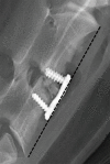
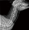
Lateral radiography of the goat neck. The length of black dot line was the distance between the upper front edge of upper vertebral body and the lower front edge of lower vertebral body.
Figure 4
Figure 4
ΔDSH of the segments after different treatments. Significant difference was not observed in the ΔDSH at 3, 6 and 12 weeks between two groups (P>0.05). ΔDSH in the cage group was significantly smaller than in the autogenous ilium group at 24 weeks (P=0.003<0.05). **, P<0.01. ΔDSH, the change of disc space height.
Figure 5
Figure 5
CT fusion scores of the segments after different treatments. (A) The fusion score in the autogenous ilium group was significantly higher than in the cage group at 12 and 24 weeks (P=0.022 and P=0.007, respectively). No significant difference was observed in the fusion score at 3 and 6 weeks between two groups. *, P<0.05; **, P<0.01; (B) in the cage group, bony junctions formed through the middle hole of the fusion cage between the upper and lower vertebral bodies, but the total fusion area was less than 30%.
Figure 6
Figure 6
The black arrow displayed the gas accumulation in the space before the vertebrae.
Figure 7
Figure 7
Volume decrease of the cages at different time points. (A) The degradation rate was relatively fast within the first 3 weeks and became slower thereafter; (B,C,D,E) the morphology of HP Mg cage on micro-CT at 3, 6, 12 and 24 weeks. HP Mg, high-purity magnesium.
Figure 8
Figure 8
Histological sections after 24 weeks after operation (scale bars =200 μm). (A) Toluidine blue staining and (B) van gieso staining showed bony junctions formed through the middle hole of the fusion device between the upper and lower vertebral bodies, and some of bone trabeculae formed continuous bone bridges which were bulky and dense; (C) HE staining and (D) Masson staining showed a 300–400 µm space between the bone tissues close to the cage, which was filled by the hyperplastic fibrous tissues. There were no evident infiltration of inflammatory cells, osteonecrosis, granuloma, and osteolysis in the space. HE, hematoxylin-eosin.
Figure 9
Figure 9
Serum biochemical parameters at different time points after treatment with the HP Mg cage. (A) The serum Mg ion concentration, (B) ALT, (C) AST, (D) CREA, and (E) UREA remained stable after surgery (all P>0.05). HP Mg, high-purity magnesium; ALT, glutamic-pyruvic transaminase; AST, glutamic oxalacetic transaminase; CREA, creatinine; UREA, urine creatinine.
Figure 10
Figure 10
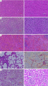
Histological findings of important tissues (HE staining) at 24 weeks after implantation. (A) Heart; (B) liver; (C) spleen; (D) lung; (E) kidney. Left scale bars =200 µm; right scale bars =100 µm. HE, hematoxylin-eosin.

Similar articles

Cited by

References

    1. Lalk M, Reifenrath J, Angrisani N, et al. Fluoride and calcium-phosphate coated sponges of the magnesium alloy AX30 as bone grafts: a comparative study in rabbits. J Mater Sci Mater Med 2013;24:417-36. 10.1007/s10856-012-4812-2 - DOI - PubMed
    1. Silber JS, Anderson DG, Daffner SD, et al. Donor site morbidity after anterior iliac crest bone harvest for single-level anterior cervical discectomy and fusion. Spine (Phila Pa 1976) 2003;28:134-9. 10.1097/00007632-200301150-00008 - DOI - PubMed
    1. Banwart JC, Asher MA, Hassanein RS. Iliac crest bone graft harvest donor site morbidity. A statistical evaluation. Spine (Phila Pa 1976) 1995;20:1055-60. 10.1097/00007632-199505000-00012 - DOI - PubMed
    1. Maldonado-Naranjo AL, Healy AT, Kalfas IH. Polyetheretherketone (PEEK) intervertebral cage as a cause of chronic systemic allergy: a case report. Spine J 2015;15:e1-3. 10.1016/j.spinee.2015.04.011 - DOI - PubMed
    1. Li ZJ, Wang Y, Xu GJ, et al. Is PEEK cage better than titanium cage in anterior cervical discectomy and fusion surgery? A meta-analysis. BMC Musculoskelet Disord 2016;17:379. 10.1186/s12891-016-1234-1 - DOI - PMC - PubMed