

In vivo retinal imaging in translational regenerative research
- PMID: 33145315
- PMCID: PMC7575995
- DOI: 10.21037/atm-20-4355
In vivo retinal imaging in translational regenerative research
Abstract
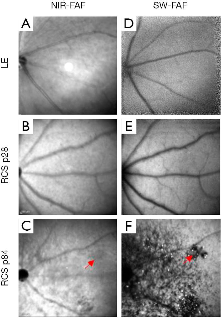
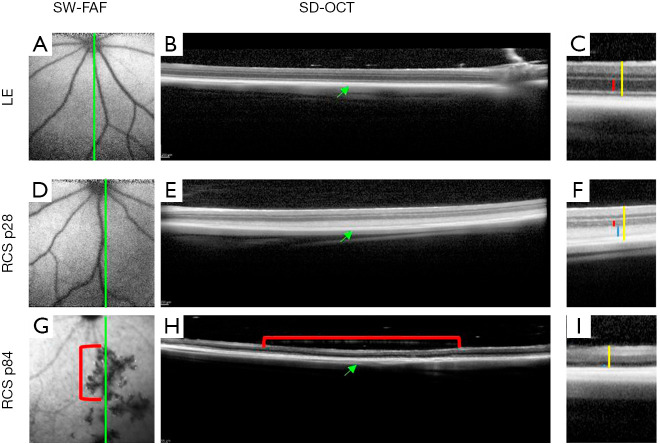
Regenerative translational studies must include a longitudinal assessment of the changes in retinal structure and function that occur as part of the natural history of the disease and those that result from the studied intervention. Traditionally, retinal structural changes have been evaluated by histological analysis which necessitates sacrificing the animals. In this review, we describe key imaging approaches such as fundus imaging, optical coherence tomography (OCT), OCT-angiography, adaptive optics (AO), and confocal scanning laser ophthalmoscopy (cSLO) that enable noninvasive, non-contact, and fast in vivo imaging of the posterior segment. These imaging technologies substantially reduce the number of animals needed and enable progression analysis and longitudinal follow-up in individual animals for accurate assessment of disease natural history, effects of interventions and acute changes. We also describe the benefits and limitations of each technology, as well as outline possible future directions that can be taken in translational retinal imaging studies.
Keywords: Retinal imaging; adaptive optics (AO); fundus imaging; optical coherence tomography (OCT).
2020 Annals of Translational Medicine. All rights reserved.
Conflict of interest statement
Conflicts of Interest: All authors have completed the ICMJE uniform disclosure form (available at http://dx.doi.org/10.21037/atm-20-4355). The series “Novel Tools and Therapies for Ocular Regeneration” was commissioned by the editorial office without any funding or sponsorship. The authors have no other conflicts of interest to declare.
Figures
Similar articles
-
Adaptive optics scanning laser ophthalmoscopy in fundus imaging, a review and update.Int J Ophthalmol. 2017 Nov 18;10(11):1751-1758. doi: 10.18240/ijo.2017.11.18. eCollection 2017. Int J Ophthalmol. 2017. PMID: 29181321 Free PMC article. Review.
-
Combined cSLO-OCT imaging as a tool in preclinical ocular toxicity testing: A comparison to standard in-vivo and pathology methods.J Pharmacol Toxicol Methods. 2020 Jul-Aug;104:106873. doi: 10.1016/j.vascn.2020.106873. Epub 2020 May 12. J Pharmacol Toxicol Methods. 2020. PMID: 32413488
-
Advances in Retinal Optical Imaging.Photonics. 2018 Jun;5(2):9. doi: 10.3390/photonics5020009. Epub 2018 Apr 27. Photonics. 2018. PMID: 31321222 Free PMC article.
-
Optical coherence tomography angiography (OCT-A) in an animal model of laser-induced choroidal neovascularization.Exp Eye Res. 2019 Jul;184:162-171. doi: 10.1016/j.exer.2019.04.002. Epub 2019 Apr 16. Exp Eye Res. 2019. PMID: 31002822
-
Review of adaptive optics OCT (AO-OCT): principles and applications for retinal imaging [Invited].Biomed Opt Express. 2017 Apr 19;8(5):2536-2562. doi: 10.1364/BOE.8.002536. eCollection 2017 May 1. Biomed Opt Express. 2017. PMID: 28663890 Free PMC article. Review.
Cited by
-
Advantages of ocular regeneration research.Ann Transl Med. 2021 Aug;9(15):1269. doi: 10.21037/atm-21-1793. Ann Transl Med. 2021. PMID: 34532406 Free PMC article. No abstract available.
References
-
- FDA. FDA approves novel gene therapy to treat patients with a rare form of inherited vision loss. Available online: https://www.fda.gov/news-events/press-announcements/fda-approves-novel-g...
-
- Machida S, Kondo M, Jamison JA, et al. P23H rhodopsin transgenic rat: correlation of retinal function with histopathology. Invest Ophthalmol Vis Sci 2000;41:3200-9. - PubMed
Publication types
LinkOut - more resources
Full Text Sources