

Do lodged foreign bodies in the neck need to be removed? No defined criteria in 2020. Fluoroscopy role and review of literature: A case report
- PMID: 33145389
- PMCID: PMC7596329
- DOI: 10.1016/j.tcr.2020.100362
Do lodged foreign bodies in the neck need to be removed? No defined criteria in 2020. Fluoroscopy role and review of literature: A case report
Erratum in
-
Erratum regarding missing Declaration of Competing Interest statements in previously published articles.Trauma Case Rep. 2023 Feb 17;45:100794. doi: 10.1016/j.tcr.2023.100794. eCollection 2023 Jun. Trauma Case Rep. 2023. PMID: 37234575 Free PMC article.
-
Erratum regarding missing patient consent statement in previously published articles.Trauma Case Rep. 2023 Mar 1;45:100809. doi: 10.1016/j.tcr.2023.100809. eCollection 2023 Jun. Trauma Case Rep. 2023. PMID: 37234577 Free PMC article.
Abstract
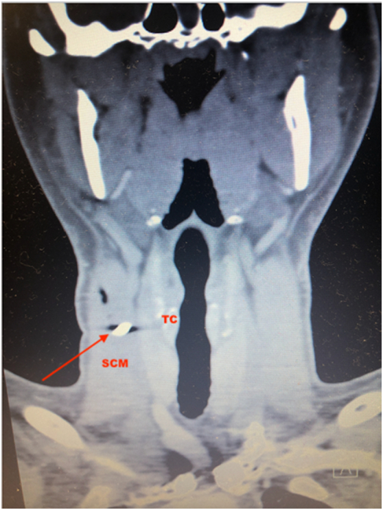
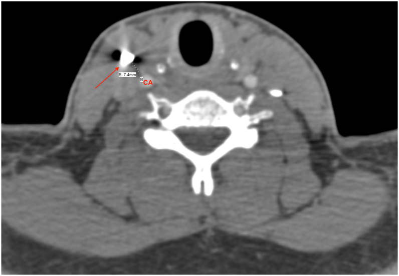
Penetrating neck wounds can be fatal and require prompt attention. The trauma literature is flooded with management protocols for penetrating wounds to the neck; however, in the absence of hard signs the definitive management of lodged foreign bodies beyond the platysma is less clear. This report describes a work-related injury of a Caucasian 33-year-old male who arrived in the Emergency Department (ER) with a 1 cm metallic foreign body (FB) lodged in zone II of the neck, 7 mm antero-lateral to the right internal carotid artery. The technical aspects of its retrieval are discussed as well as a literature review of the current management of embedded FBs in the neck. The patient was taken to the operating room and the FB was removed via a 3 cm incision. Fluoroscopy was used for exact localization of and to allow a precise skin incision overlying the FB. The FB was retrieved uneventfully; a fiberoptic esophagoscopy and bronchoscopy showed normal findings. The patient was discharged home the next day. At 15 months follow-up he is doing well without sequela. The use of fluoroscopy is strongly encouraged for FB removal in asymptomatic patients. The management of lodged foreign bodies in the neck should be part of future guidelines.
Keywords: Fluoroscopy; Foreign body; Neck trauma; Removal.
© 2020 The Author.
Figures


References
-
- American College of Surgeons National Trauma Data Bank Annual Report. 2013. http://www.facs.org/trauma/ntdb/pdf/ntdb-annual-report-2013.pdf [Accessibility verified May 1, 2014]
-
- Wang S., Liu J., Chen Y., Yang X., Xie D., Li S. Diagnosis and treatment of nine cases with carotid artery rupture due to hypopharyngeal and cervical esophageal foreign body ingestion. Eur. Arch. Otorhinolaryngol. 2013;270:1125–1130. - PubMed
-
- Rodriguez Gomez E. Cervical foreign body. An unusual case. Acta Otorinolaringol. Esp. 2004;55(6):298–301. - PubMed
Publication types
LinkOut - more resources
Full Text Sources
